 X-ray imaging is a broad term that covers several types of studies that require the visualization of the internal parts of the body using x-ray techniques.
It is used to diagnose or treat patients by visualizing internal structure of the body to assess the presence or absence of disease, foreign objects, and structural damage or anomaly.
During a radiographic procedure, an x-ray beam is passed through the body. A portion of the x-rays are absorbed or scattered by the internal structure and the remaining x-ray pattern is transmitted to a detector so that an image may be recorded for later evaluation. The recoding of the pattern may occur on film or through electronic means.
Uses
X-ray imaging has many applications in medical diagnostics, used to visualize various body structures. The following are the most used applications:
· Radiography
· Bone mineral density scan
· Mammography
· Fluoroscopy
· CT scanning
Risks/Benefits
The radiation dose the patient receives varies depending on the individual procedure, but is generally less than that received during fluoroscopy and computed tomography (CT) procedures.
The major risks associated with x-ray imaging are the small possibilities of:
· developing a radiation-induced cancer or cataracts some time later in life, and
· causing a disturbance in the growth or development of an embryo or fetus (teratogenic defect) when performed on a pregnant patient or one of childbearing age.
When an individual has a medical need, the benefit of radiography far exceeds the small cancer risk associated with the procedure. Even when radiography is medically necessary, it should use the lowest possible exposure and the minimum number of images. In most cases many of the possible risks can be reduced or eliminated with proper shielding.
Applicable Tests
Abdomen KUB
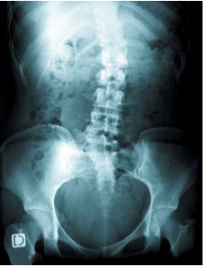
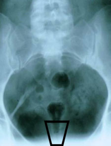
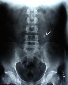 Radiography (X-Ray) is the use of ionizing electromagnetic radiation such as X-rays to view anatomy of various body structures. KUB stands for Kidneys, Ureters, and Bladder.
A KUB X-Ray is a plain frontal supine radiograph of the abdomen. It is often supplemented by an upright PA view of the chest (to rule out air under the diaphragm or thoracic etiologies presenting as abdominal complaints) and a standing view of the abdomen (to differentiate obstruction from ileus by examining gastrointestinal air/water levels).
-
How the test is done
You will be asked to lie on your back on the x-ray table. The x-ray machine is positioned over your abdominal area. The technician will ask you to hold your breath when the picture is taken so that the picture is not be blurry. You may be asked to change position to the side or to stand up for additional pictures. There is no discomfort while this procedure. The test usually takes about 15 minutes.
-
How to prepare for the test
Before having the x-ray, tell the health care provider the following:
· If you are pregnant or think you could be pregnant
· Have an IUD inserted
· Have had a barium contrast media x-ray in the last 4 days
· If you have taken any medicines such as Pepto Bismol in the last 4 days (this type of medicine can interfere with the x-ray)You must remove all the jewelry.
-
Why the test is done
· Diagnose a pain in the abdomen or unexplained nausea
· Identify suspected problems in the urinary system, such as a kidney stone
· Identify blockage in the intestine
· Locate an object that has been swallowed -
What abnormal results mean
Abnormal findings include:
· Abdominal masses
· Buildup of fluid in the abdomen
· Certain types of gallstones
· Foreign object in the intestines
· Hole in the stomach or intestines
· Injury to the abdominal tissue
· Intestinal blockage
· Kidney stones -
Risks
There is a low radiation exposure. X-rays are monitored and regulated to provide the minimum amount of radiation exposure needed to produce the image. Most experts feel that the risk is low compared with the benefits.
Pregnant women and children are more sensitive to the risks of an x-ray. Women should tell the health care provider if they are, or may be, pregnant.
-
Considerations
The test is not usually recommended for pregnant women. The ovaries and uterus cannot be shielded during the abdominal x-ray because of their location.
Men should have a lead shield placed over the testes to protect against the radiation.
Neck soft tissues
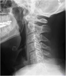 A neck X-ray (also called a cervical spine X-ray) is an X-ray image taken of your cervical vertebrae, which are the seven bones of your neck that encase and protect the top section of your spinal cord. A neck X-ray also shows the surrounding structures, including your vocal cords, tonsils, adenoids, trachea (windpipe), and epiglottis (the flap of tissue that covers your windpipe when you swallow).
An X-ray is a form of radiation that passes through your body to expose a piece of film, forming an image of your body. Dense structures, like bones, appear white on X-rays because very little radiation can pass through them to expose the film on the other side.
Soft tissues, such as blood vessels, skin, fat, and muscles, are less dense, so more radiation can pass through them. These structures will appear dark gray on the X-ray image.
-
Why is a neck x-ray performed?
If you have a neck injury or persistent numbness, pain, or weakness in your upper extremities, your doctor may request an X-ray. Your doctor will check the X-ray image for evidence of the following conditions:
· fractured or broken bones
· swelling in or near your airway
· thinning of the neck bones due to osteoporosis
· bone tumors or cysts
· chronic wear on the disks and joints of your neck (cervical spondylosis)
· joints that are pushed out of their normal positions (dislocations)
· abnormal growths on the bones (bone spurs)
· spinal deformities
· swelling around the vocal cords (croup)
· inflammation of the tissue that covers your windpipe (epiglottitis)
· a foreign object that is lodged in your throat or airway
· enlarged tonsils and adenoids -
What are the risks of a neck x-ray?
X-rays are very safe and generally produce no side effects or complications. The amount of radiation used in a single X-ray is quite small but should not be over used.
Children and unborn babies are especially sensitive to radiation, so tell your doctor if you are pregnant so that precautions can be taken during the procedure. If you are pregnant and must have a neck X-ray, you will be given a lead vest to cover your abdomen to keep radiation from harming your fetus. Children will also be given a lead shield to cover their abdomens to protect their reproductive organs from the radiation.
-
How is a neck x-ray performed?
The technician will ask you to remove any clothing and jewelry on your upper body, because metal can interfere with the X-ray equipment.
You will be asked to lie down flat on the X-ray table and the X-ray machine will be moved over your neck area. You must be very still and hold your breath for a few moments while the image is taken so that it won’t be blurry. The radiology tech will probably ask you to lie in several different positions so that the X-ray can be taken from multiple angles. The procedure is painless and generally takes 15 minutes or less.
-
What do the test results mean?
The radiology technologist will develop the X-rays and send them to your doctor within a few days. If your bones and tissues appear normal on the X-ray images, you probably do not have bone spurs, spinal deformities, cervical spondylosis, etc. If any of these abnormalities appear on your X-rays, your doctor will discuss your treatment options with you.
Skull
 A skull X-ray is an imaging test used to see the bones of the skull, including the facial bones, the nose, and the sinuses. It is an easy, quick, and effective method that has been used for decades to help doctors view the area that houses your most vital organ—brain.
-
Why a skull x-ray is done?
A skull X-ray is typically done after a traumatic head injury. The X-ray allows doctors to inspect any damage from the injury. Other reasons you may undergo a skull X-ray include:
· decalcification of the bone
· deformities in the skull
· fractures
· frequent headaches
· infection of the bones of the skulls
· occupational hearing loss
· tumorsPrior to your X-ray, your doctor will tell you the exact reason for your X-ray.
-
The risks of a skull x-ray
While X-rays use radiation, none of it remains in your body when the test is done. Doctors argue that the benefits of the test outweigh any risk from exposure to the minimal amount of radiation produced.
However, while the level of exposure is considered safe for adults, it is not safe for developing fetuses. If you are pregnant or believe you are pregnant, tell your doctor, as the X-ray may be unsafe for your unborn child.
-
How to prepare for a skull x-ray
X-rays require little preparation on the patient’s part. You will have to remove any jewelry, eyeglasses, and other metals from around your head. This includes necklaces and earrings. Metal can interfere with the clarity of the X-ray image.
Inform your doctor if you have any kind of surgically implanted device, such as a metal plate in your head, an artificial heart valve, or a pacemaker. Even though these things might interfere somewhat with the image, your doctor may still choose to use X-ray.
-
How a skull x-ray is performed
The X-ray will be performed in a special room with a movable X-ray camera attached to a large metal arm. It’s designed to be able to take multiple X-rays of various body parts.
For a skull X-ray, you’ll sit in a chair or lie down on a specialized table. A lead apron may be placed over your body, especially your pelvic region and breasts, to protect from radiation.
The X-ray technician may have you lie on your back to start, but you’ll have to change positions so the camera can capture front and side views. While the images are being taken, you’ll be asked to hold your breath and stay very still. You won’t feel the X-ray pass through you.
After the right images have been captured—which should only take about 20 minutes or so—the test is finished.
Sinus
 A sinus X-ray uses radiation to form a picture of your sinuses. There are four different sets of sinus passages, which are the air-filled spaces in the front of the face: over and around the eyes, in the cheekbones, between the eyes, and behind the eyes.
The passages will look black on an X-ray of healthy sinuses. This is because they are filled with air, which appears black on X-rays. A grey or white area on the X-ray of the sinuses indicates a problem. Most often, this is due to fluid or inflammation in the sinuses.
A sinus X-ray may also be called X-ray of the sinuses or paranasal sinus radiography. Sinus X-rays are a noninvasive test that can be completed quickly and with little discomfort or pain.
-
What does a sinus x-ray diagnose?
Your doctor will order a sinus X-ray when you are experiencing symptoms of sinus problems or sinusitis, also known as a sinus infection.
Symptoms of sinusitis include:· stuffy nose with thick nasal secretions that may appear white, yellow, or green
· pain or tenderness in the forehead, between the eyes, in the cheeks, upper jaw, and teeth
· feeling of fullness in the face and swelling around the eyes, around the nose, and in the cheeks
· decreased sense of smell
· fatigue and general feeling of being unwell (malaise)
· cough, which is typically worse at night
· sore throat
· earache
· feverSinusitis can either be acute or chronic. Acute sinusitis typically lasts a couple of weeks and can be caused by infectious or non-infectious causes. Infections that can cause acute sinusitis that would include viral infections (including the common cold), fungal infections, and bacterial infections. Additionally, anything that raises your risk of getting a sinus infection can cause acute sinusitis. Sinusitis can be brought about by non-infectious causes as well, such as allergies, reduced immune function, enlarged or infected adenoids in children, and tumors or polyps in the nasal passages or sinuses.
Chronic sinusitis means that the sinuses are inflamed and infected for 12 weeks or more. This can be caused by many of the same things that cause acute sinusitis.Causes for chronic sinusitis include infection, asthma, allergies, recurring acute sinusitis, trauma to the face, problems with the respiratory tract, and immune deficiency disorders.
A sinus X-ray may also be used to diagnose other sinus problems, including hemorrhage (bleeding) or tumor in the sinuses. This imaging can also be used to help diagnose meningitis (infection in the brain and spinal cord) or orbital cellulitis (an infection around the eye).
-
What happens during a sinus x-ray?
There is no preparation required for the procedure, except for removing any jewelry or metal objects you may be wearing. A radiologist or X-ray technician will perform the sinus X-ray.
You may be seated or asked to lie down on an X-ray table. The radiologist will place a lead apron over your torso to help protect you from radiation. The radiologist will then place your head in line with the X-ray machine. You will need to hold this position as the X-ray technician steps behind a protective window to take the X-ray.
It is important to remain still as possible while the X-ray is being taken, otherwise the image will be blurred. It will only take a couple of seconds to take the X-ray, during which time you may hear a clicking sound, similar to the sound a camera makes when taking a picture.
The technologist may need to reposition you several times in order to get images of all the sinuses.
-
What are the risks of a sinus x-ray?
An X-ray uses radiation to take images of the body. While an X-ray uses relatively low amounts of radiation, there is still a risk every time the body is exposed to radiation. It is important that your doctor knows what medical tests you have had in the past, so you are not overexposed to radiation.
If you are pregnant or think you might be pregnant, it important to inform your doctor, as radiation can cause birth defects. Your doctor may wish to order a different test, or use special measures to protect the fetus from radiation.
-
Further testing for sinus issues
Sinus X-rays are less invasive than other sinus tests, but they’re also less comprehensive. While sinus X-rays can only be used to indicate the presence of a problem, other sinus tests can be used to help determine the cause of a sinus problem.
These tests include:
· nasal endoscopy or rhinoscopy
· blood tests
· computed tomography (CT) scan
· magnetic resonance imaging (MRI)
· sinus puncture and bacteria culture
Orbits X-Ray
 Orbital x-ray is a low-radiation imaging study of an area and structures containing the eye. The orbit is the circle of thin bones that houses and protects the eye, even extending behind the eye and nearly wrapping around it. The orbit includes the eyebrow, the bridge of the nose and the cheekbone.
Orbital x-ray, or orbital radiography, is often used to detect problems resulting from injury or trauma to the eye. The exam may also detect changes to the structure of the eye, which may indicate various diseases. An ophthalmologist may also order an orbital x-ray if there is concern that foreign bodies may be present in the eye that cannot be detected with an instrument called an ophthalmoscope.
-
Precautions
Pregnant women and women who could possibly be pregnant should only receive orbital x-rays when absolutely necessary. If the patient is in severe pain due to injury or trauma, a painkiller may be given to help ease discomfort during positioning of the head throughout the exam. No other precautions are necessary for orbital x-rays.
-
How the procedure is done
The orbital x-ray involves several different views in order for the physician to clearly see various parts of the eye without obstruction. In orbital x-rays, images of the unaffected eye may also be taken to compare its shapes and structures to those of the affected eye. Views may include side view (lateral), back to front (posteroanterior), base view, views from both sides, and an image from the center to one outside edge (half-axial projection). Projections of the optical canal will also be included. For all of these views, the patient may be seated upright or asked to lie on a table in the x-ray room.
The orbital x-ray procedure should take about 15 minutes to complete. Following the procedure, the patient will usually be asked to wait until the pictures are processed to ensure they are high enough quality and that repeat x-rays are not necessary.
-
Preparation
There are no special dietary preparations needed prior to an orbital x ray. As with any radiography procedure, the patient should remove any jewelry or metal objects, which may interfere with a clear image.
-
Risks
Radiation exposure is low for this procedure and all certified radiology facilities follow strict personnel and equipment guidelines for radiation protection. Women of child bearing age and children should be offered protective shielding (lead aprons) to cover the genital and/or abdominal areas.
-
Normal results
Normal findings will show the bones of the orbit intact, and will show similarity between the orbit that is being studied and the unaffected orbit.
-
Abnormal results
Positive findings from an orbital x-ray may show that there has been injury to the eye. Certain signs may indicate some disease that is affecting the orbital structures. Tiny fractures in the orbital bones can usually be detected on the radiograph. The floor bone, the medial wall and the ethmoid bone, which is a spongy bone that forms the upper part of the nasal cavity, are the most likely to break. In a blowout fracture (one involving the orbital floor), radiographic findings may include disruption to the orbital floor, an opaque look to the sinuses on the same side as the affected orbit (due to hemorrhage) or signs of sinus problems from the orbital root’s interference. These indications can be seen in most typical orbital x-ray views.
Since the physician examines both orbits side by side, indications of differences in size and shape of the various structures in the orbit may be apparent. The orbit may be enlarged, indicating irritation from an injury or foreign body. A number of growing tumors within the eye or brain area may also cause orbital enlargement. Destruction of the walls of the orbit may indicate a nearby infection or malignancy. Changes in density of the tiny orbit bones may also be a sign of bone disease or cancer spread to bone.
Children’s orbits are more likely to be enlarged by a fast growing lesion, since their orbital bones have not fully developed.
Chest
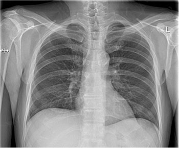 Chest radiograph, colloquially called a chest X-ray (CXR) is a projection radiograph of the chest used to diagnose conditions affecting the chest, its contents, and nearby structures. Chest radiographs are among the most common films taken, being diagnostic of many conditions.
Like all methods of radiography, chest radiography employs ionizing radiation in the form of X-rays to generate images of the chest. The mean radiation dose to an adult from a chest radiograph is around 0.02 mSv (2 mrem) for a front view (PA or posterior-anterior) and 0.08 mSv (8 mrem) for a side view (LL or latero-lateral).
-
Conditions commonly identified by chest radiography
· Pneumonia
· Pneumothorax
· Interstitial lung disease
· Congestive heart failure
· Bone fracture
· Hiatal herniaChest radiographs are used to diagnose many conditions involving the chest wall, including its bones, and also structures contained within the thoracic cavity including the lungs, heart, and great vessels. Pneumonia and congestive heart failure are very commonly diagnosed by chest radiograph.
For some conditions of the chest, radiography is good for screening but poor for diagnosis. When a condition is suspected based on chest radiography, additional imaging of the chest can be obtained to definitively diagnose the condition or to provide evidence in favor of the diagnosis suggested by initial chest radiography. Unless a fractured rib is suspected of being displaced, and therefore likely to cause damage to the lungs and other tissue structures, x-ray of the chest is not necessary as it will not alter patient management.
The main regions where a chest X-ray may identify problems may be summarized as ABCDEF by their first letters:
· Airways, including hilar adenopathy or enlargement
· Breast shadows
· Bones, e.g. rib fractures and lytic bone lesions
· Cardiac silhoutte, detecting cardiac enlargement
· Costophrenic angles, including pleural effusions
· Diaphragm, e.g. evidence of free air, indicative of perforation of an abdominal viscus
· Edges, e.g. apices for fibrosis, pneumothorax, pleural thickening or plaques
· Extrathoracic tissues
· Fields (lung parenchyma), being evidence of alveolar flooding
· Failure, e.g. alveolar air space disease with prominent vascularity with or without pleural effusions -
Views
PA chest x-ray
Different views (also known as projections) of the chest can be obtained by changing the relative orientation of the body and the direction of the x-ray beam. The most common views are posteroanterior, anteroposterior, and lateral. In a posteroanterior (PA) view, the x-ray source is positioned so that the x-ray beam enters through the posterior (back) aspect of the chest, and exits out of the anterior (front) aspect where the beam is detected. To obtain this view, the patient stands facing a flat surface behind which is an x-ray detector. A radiation source is positioned behind the patient at a standard distance (most often 6 feet, 1,8m), and the x-ray beam is fired toward the patient.
AP chest x-ray
In anteroposterior (AP) views, the positions of the x-ray source and detector are reversed: the x-ray beam enters through the anterior aspect and exits through the posterior aspect of the chest. AP chest x-rays are harder to read than PA x-rays and are therefore generally reserved for situations where it is difficult for the patient to get an ordinary chest x-ray, such as when the patient is bedridden. In this situation, mobile X-ray equipment is used to obtain a lying down chest x-ray (known as a “supine film”). As a result, most supine films are also AP.
A chest radiograph with the angle parts of the ribs
Lateral views of the chest are obtained in a similar fashion as the posteroanterior views, except in the lateral view, the patient stands with both arms raised and the left side of the chest pressed against a flat surface.
Spine
 Spinal X-rays are pictures of the spine. They may be taken to find injuries or diseases that affect the discs or joints in your spine. These problems may include spinal fractures, infections, dislocations, tumors, bone spurs, or disc disease.
Spinal X-rays are also done to check the curve of your spine (scoliosis) or for spinal defects.
The spine is divided into four parts. So there are four common types of spinal X-rays:
 Cervical spine X-ray: This X-ray test takes pictures of the 7 neck (cervical) bones.
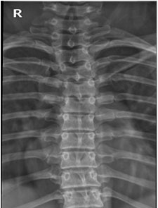 Thoracic spine X-ray: This X-ray test takes pictures of the 12 chest (thoracic) bones.
 Lumbosacral spine X-ray: This X-ray test takes pictures of the 5 bones of the lower back (lumbar vertebrae) and a view of the 5 fused bones at the bottom of the spine (sacrum).
 Sacrum/coccyx X-ray: This X-ray test takes a detailed view of the 5 fused bones at the bottom of the spine (sacrum) and the 4 small bones of the tailbone (coccyx).
The most common spinal X-rays are of the cervical vertebrae (C-spine films) and lumbosacral vertebrae (LS-spine films).
-
Why is it done?
A spinal X-ray is done to:
· Find the cause of ongoing pain, numbness, or weakness.
· Check for arthritis of the joints between the vertebrae and the breakdown (degeneration) of the discs between the spinal bones.
· Check injuries to the spine, such as fractures or dislocations.
· Check the spine for effects from other problems, such as infections, tumors, or bone spurs.
· Check for abnormal curves of the spine, such as scoliosis, in children or young adults.
· Check the spine for problems present at birth (congenital conditions), such as spina bifida, in infants, children, or young adults.
· Check changes in the spine after spinal surgery. -
How to prepare
Before the X-ray test, tell your doctor if you are or might be pregnant. The risk of radiation exposure to your unborn baby (fetus) must be considered. The risk of damage from the X-rays is usually very low compared with the potential benefits of the test. If a spinal X-ray is absolutely necessary, a lead apron will be placed over your belly to shield your baby from the X-rays.
You may need to take off any jewelry that may be in the way of the X-ray picture, such as if you have a pierced belly button.
You don’t need to do anything else before you have this test.
Talk to your doctor about any concerns you have regarding the need for the test, its risks, how it will be done, or what the results will mean.
-
How is it done?
A spinal X-ray is taken by a radiology technologist. The X-ray pictures are usually read by a doctor who specializes in reading X-rays (radiologist).
You will need to remove any jewelry that may be in the way of the X-ray picture. You may need to take off some of your clothes, depending on which area is examined. You will be given a cloth or paper gown to use during the test. You may be allowed to keep on your underwear if it does not get in the way of the test.
During the X-ray test, you will lie on an X-ray table. If the X-ray is being taken because of a possibly serious injury to your neck or back, to
prevent causing more injury a radiologist will look at the first X-ray pictures before taking others. If you have a neck brace (cervical collar) in place, X-ray pictures may be taken and a physical exam done to see whether the brace can be taken off without hurting the spine.Usually 3 to 5 X-ray pictures are taken. You need to lie very still to avoid blurring the pictures.
A spinal X-ray usually takes about 15 minutes.
-
How it feels
You will feel no discomfort from the X-rays.
-
Risks
There is always a slight risk of damage to cells or tissue from being exposed to any radiation, including the low levels of radiation used for this test. But the risk of damage from the X-rays is usually very low compared with the potential benefits of the test.
For example, the radiation exposure from a chest X-ray is about equal to the natural radiation exposure received during a round-trip airline flight from Boston to Los Angeles (or Montreal to Vancouver) or 10 days in the Rocky Mountains (Denver, Colorado).
-
Results
In an emergency, a doctor can see the results of a spinal X-ray in a few minutes. Otherwise, a radiologist usually has the official X-ray report ready the next day.
Spinal X-ray:
Normal:
· The bones of the spine (vertebrae) are normal in number, size, shape, appearance, and how they are lined up.
· No broken bones, dislocations, or foreign objects are present. The soft tissues around the vertebrae look normal.
· The spine is not abnormally curved.Abnormal:
· Broken bones, dislocations, or foreign objects are present.
· The spine is abnormally curved, such as from scoliosis.
· Diseases that affect the spine, such as thin bones (osteoporosis) or arthritis, are present. One or more bones in the spine may be abnormal because of a condition you were born with or because of cancer, infection, or trauma.
· Disc disease, which is fairly common, can sometimes be seen on a spinal X-ray as a narrowed space between the bones of the spine. Bone spurs can also be seen. -
What affects the test
Reasons you may not be able to have the test or why the results may not be helpful include:
· If you are pregnant. The X-rays may not be safe for the fetus.
· If you can’t remain still during the test. The pictures may not be clear.
· If you are very overweight. This can make it hard to see the details of the spinal X-ray. -
What to think about
Your X-ray results may be different from earlier test results because you were tested at a different medical center or you had a different kind of test.
The most common causes of low back pain, such as strained back muscles or ligaments, do not show up on a spinal X-ray.
Other tests, such as a CT scan, an MRI, or a myelogram, provide more information about the spinal bones, joints, nerves, discs, muscles, and ligaments than a spinal X-ray.
Pelvis X-ray
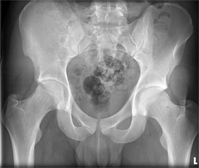 An X-ray is a common imaging test that has been used for decades to help doctors view the inside of the body without having to make an incision.
A pelvis X-ray looks specifically at your pelvic region—the area between your hips that holds many of your reproductive and digestive organs. Your pelvis is actually made up of three bones—the ilium, ischium, and pubis—and also forms your hip joint.
Like all X-rays, this test uses a small amount of radiation, so it may not be recommended for pregnant women or small children.
-
Why a pelvis x-ray is done
Your doctor may order a pelvic X-ray for numerous reasons. Often, an X-ray is taken after a traumatic event, such as a car accident or a fall.
Pelvic X-ray can help your doctor detect various conditions, such as:
· arthritis that affects your hip
· inflammation where your sacrum joins the ilium (sacroilitis)
· pelvic fractures
· stiffness of the spine or sacroiliac joint (ankylosing spondylitis)
· tumors (NIH ) -
Risks of a pelvis x-ray
X-rays use small amounts of radiation. The level of exposure is considered safe for adults. However, it is not considered safe for developing fetuses. If you are pregnant or believe you could be pregnant, tell your doctor before the procedure. He or she may suggest alternative testing methods that do not use radiation, such as a magnetic resonance imaging (MRI) scan.
If you are having an X-ray due to a traumatic event that caused pain and possibly a broken pelvis, you may experience additional pain during the X-ray. The test requires you to adjust your body so that clear images can be taken. This may cause some discomfort. If you are worried, you can ask your doctor for pain medicine prior to your X-ray.
For some X-rays, your doctor will inject you with a contrast dye before the procedure in order to improve the images. The dye—usually iodine—can cause some side effects. These include hives, itching, lightheadedness, nausea, or a metallic taste in the mouth. In very rare cases, the dye can cause a severe reaction, such as anaphylactic shock, very low blood pressure, or cardiac arrest.
-
How to prepare for a pelvis x-ray
X-rays are standard procedures and involve almost no preparation from the patient.
Depending on the area under review, you may want to wear loose, comfortable clothing that you can easily move around in. You may also be asked to change into a gown for the test.
You will be instructed to remove any jewelry and other metallic items from your body before the X-ray is taken. You should always tell your doctor if you have any metal implants from prior surgeries. These can block the X-rays from passing through your body.
If your test requires contrast dye, a doctor or nurse will give it to you as an injection, an enema, or a pill to swallow before the test.
-
How a pelvis x-ray is performed
X-rays are performed in a hospital’s radiology department or a clinic that specializes in diagnostic procedures. Once you are fully prepared, an X-ray technician—called a radiologist—will tell you how he or she needs you to be positioned in order to get the right view.
The technician will most likely require you to lie, sit, or stand in several positions during the test. Some images may be taken while you stand in front of a specialized plate that contains X-ray sensors.
In some cases, the technician will move a large camera connected to a steel arm over your body. This can capture the X-ray images of your body using film or sensors held in the table.
While the images are being taken, you will have to hold your breath and remain still. This provides the clearest images possible.
The test is finished usually in 15 minutes.
X-Ray of Extremities
 X-rays are made by using external radiation to produce images of the body, its organs, and other internal structures for diagnostic purposes.
When the body undergoes X-rays, different parts of the body allow varying amounts of the X-ray beams to pass through. Images are produced in degrees of light and dark, depending on the amount of X-rays that penetrate the tissues. The soft tissues in the body (such as blood, skin, fat, and muscle) allow most of the X-ray to pass through and appear dark gray on the film. A bone or a tumor, which is denser than soft tissue, allows few of the X-rays to pass through and appears white on the X-ray. At a break in a bone, the X-ray beam passes through the broken area and appears as a dark line in the white bone.
X-rays of the extremities are often used as the first step in diagnosing injuries of the extremities, but may also be used to evaluate other problems involving the bones and/or soft tissues.
Other related procedures that may be used to diagnose problems involving the extremities include computed tomography (CT scan), magnetic resonance imaging (MRI), arthrography, or bone scan.
-
Reasons for the procedure
X-rays of the extremities (such as the arm, leg, hand, foot, ankle, shoulder, knee, hip or hand) may be performed to assess the bones of the extremity for injuries, such as fractures or broken bones, or evidence of other injuries or conditions, such as infection, arthritis, tendinitis, bone spurs, tumors, or congenital abnormalities. X-rays of the extremities may also be used to evaluate bone growth and development in children.
X-rays of joints may be done to evaluate damage to soft tissues, such as cartilage, muscle, tendons, or ligaments, and to assess for the presence of fluid in the joint, and other abnormalities of the joint such as bone spurs, narrowing of the joint, and changes in the structure of the joint.
There may be other reasons for your doctor to recommend an X-ray of the extremities.
-
Risks of the procedure
You may want to ask your doctor about the amount of radiation used during the procedure and the risks related to your particular situation. It is a good idea to keep a record of your past history of radiation exposure, such as previous scans and other types of X-rays, so that you can inform your doctor. Risks associated with radiation exposure may be related to the cumulative number of X-ray examinations and/or treatments over a long period of time.
If you are pregnant or suspect that you may be pregnant, you should notify your doctor. Radiation exposure during pregnancy may lead to birth defects. If it is necessary for you to have an X-ray of the extremities, special precautions will be made to minimize the radiation exposure to the fetus.
There may be other risks depending on your specific medical condition. Be sure to discuss any concerns with your doctor prior to the procedure.
-
Before the procedure
Your doctor will explain the procedure to you and offer you the opportunity to ask questions that you might have about the procedure.
Generally, no prior preparation, such as fasting or sedation, is required.
Notify the technologist if you are pregnant or suspect you may be pregnant.
Notify the technologist if you have had a recent barium X-ray procedure, as this may interfere with obtaining an optimal X-ray exposure of the lower back area during a hip X-ray.Based on your medical condition, your doctor may request other specific preparation.
-
During the procedure
Generally, an X-ray procedure of the extremities follows this process:
You will be asked to remove any clothing, jewelry, hairpins, eyeglasses, hearing aids, or other metal objects that might interfere with the procedure.
If you are asked to remove clothing, you will be given a gown to wear.
The type of procedure being performed will dictate your positioning, such as lying on a table, sitting, or standing, and the type of X-ray equipment used. You will be positioned on an X-ray table that carefully places the part of the body that is to be X-rayed between the X-ray machine and a cassette containing the X-ray film or digital media. Examinations in the sitting or standing position are performed in a similar manner, with the body part being examined placed between the X-ray machine and the X-ray digital media.
Body parts not being imaged may be covered with a lead apron (shield) to avoid exposure to the X-rays.
The technologist will ask you to hold the extremity still in a certain position for a few moments while the X-ray exposure is made.If the X-ray is being performed to determine an injury, special care will be taken to prevent further injury. For example, a splint or brace may be applied to the leg or arm if a fracture is suspected.
Some X-ray studies may require several different positions of the extremity. It is extremely important to remain completely still while the exposure is made, as any movement may distort the image and even require another X-ray to be done to obtain a clear image of the body part in question.
While the X-ray procedure itself causes no pain, the manipulation of the body part being examined may cause some discomfort or pain, particularly in the case of a recent injury or invasive procedure such as surgery. The technologist will use all possible comfort measures and complete the procedure as quickly as possible to minimize any discomfort or pain.