Radiation Dose in X-Ray, CT Exams and Nuclear Imaging
What are x-rays and what do they do?
X-rays are forms of radiant energy, like light or radio waves. Unlike light, x-rays can penetrate the body, which allows a radiologist to produce pictures of internal structures. The radiologist can view these on computer monitor.
X-ray examinations provide valuable information about your health and play an important role in helping your doctor make an accurate diagnosis. In some cases x-rays are used to assist with the placement of tubes or other devices in the body or with other therapeutic procedures.
Measuring radiation dosage
The scientific unit of measurement for radiation dose, commonly referred to as effective dose, is the millisievert (mSv). Other radiation dose measurement units include rad, rem, roentgen, sievert, and gray.
Because different tissues and organs have varying sensitivity to radiation exposure, the actual radiation risk to different parts of the body from an x-ray procedure varies. The term effective dose is used when referring to the radiation risk averaged over the entire body.
The effective dose accounts for the relative sensitivities of the different tissues exposed. More importantly, it allows for quantification of risk and comparison to more familiar sources of exposure that range from natural background radiation to radiographic medical procedures.
Vitalim assures that amount of radiation you are exposed would always be As Low As Reasonably Achievable in order to get a quality image but not to put the patient at risk.
Naturally-occurring “background” radiation exposure
We are exposed to radiation from natural sources all the time. According to recent estimates, the average person in North America receives an effective dose of about 3 mSv per year from naturally occurring radioactive materials and cosmic radiation from outer space. These natural “background” doses vary throughout the country.
People living in the plateaus of Colorado or New Mexico receive about 1.5 mSv more per year than those living near sea level. The added dose from cosmic rays during a coast-to-coast round trip flight in a commercial airplane is about 0.03 mSv. Altitude plays a big role, but the largest source of background radiation comes from radon gas in our homes (about 2 mSv per year). Like other sources of background radiation, exposure to radon varies widely from one part of the country to another.
To explain it in simple terms, we can compare the radiation exposure from one chest x-ray as equivalent to the amount of radiation exposure one experiences from our natural surroundings in 10 days.
Following are comparisons of effective radiation dose with background radiation exposure for several radiological procedures described within this website:
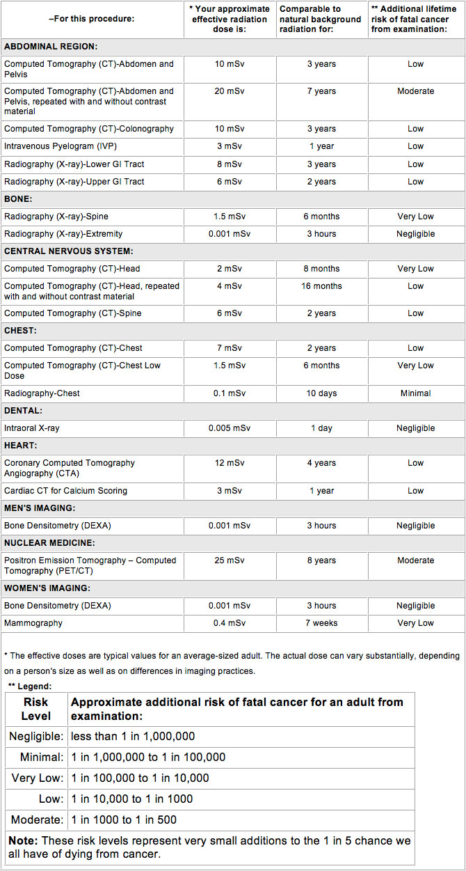
Please note that the above chart attempts to simplify a highly complex topic for patients’ informational use. The effective dose listed above may be used to estimate cancer and cancer related deaths.
X-ray safety
As with other medical procedures, x-rays are safe when used with care. Radiologists and x-ray technologists have been trained to use the minimum amount of radiation necessary to obtain the needed results. Properly conducted imaging carries minimal risks and should be performed when clinically indicated. The amount of radiation used in most examinations is very small and the benefits greatly outweigh the risk of harm.
X-rays are produced only when a switch is momentarily turned on. As with visible light, no radiation remains after the switch is turned off.
X-rays over your lifetime
The decision to have an x-ray exam is a medical one, based on the likelihood of benefit from the exam and the potential risk from radiation. For low dose examinations, usually those that involve only films taken by a technologist, this is generally an easy decision. For higher dose exams such as computed tomography (CT) scans and those involving the use of contrast materials (dyes) such as barium or iodine, the radiologist may want to consider your past history of exposure to x-rays. If you have had frequent x-ray exams and change healthcare providers, it is a good idea to keep a record of your x-ray history for yourself. This can help your doctor make an informed decision. It is also very important to tell your doctor if you are pregnant before having an exam that involves the abdomen or pelvic region.
Pregnancy and x-rays
As with any aspect of medical care, knowing that a patient is or could be pregnant is important information. Pregnancy, for example, might explain certain symptoms or medical findings. When a pregnant patient is ill or injured, the physician will carefully select medications to avoid potential risks to the developing child. This is also true of x-rays.
While the vast majority of medical x-rays do not pose a critical risk to a developing child, there may be a small likelihood of causing a serious illness or other complication. The actual risk depends on how far along the pregnancy is and on the type of x-ray. Ultrasound studies, for example, don’t use x-rays and have never demonstrated any potential risk to pregnancy. X-ray studies of the head, arms, legs and chest do not usually expose the baby directly to x-rays and typically the technologist who takes the x-rays will implement special precautions to ensure that the baby of a pregnant patient is not directly exposed.
Sometimes patients need examinations of the abdomen or pelvis while they are pregnant. When studies of the abdomen or pelvis are required, the physician may prefer to order a different type of exam for a pregnant patient or reduce the number of x-rays from that which is normally acquired. Therefore, it is important that you inform your physician or the x-ray technologist about your reproductive status before the x-ray study is performed.
Most standard x-ray examinations of the abdomen are not likely to pose a serious risk to the child. Some abdominal and pelvic studies such as CT deliver greater amounts of radiation to a developing pregnancy. Informing the radiologist that you are or might be pregnant is important so that your medical care can be planned with both you and your baby in mind. Remember, this is done to optimize medical care by reducing any potential risk.
Radionuclide exams, also known as nuclear medicine, use an x-ray-like radiation. The method of use, however, is quite different from x-rays and produces very different looking images. Please inform your physician or the nuclear medicine technologist about any possible pregnancy or breast feeding before the examination begins.
Some of the pharmaceuticals that are used for the study can pass into the mother’s milk and subsequently the child will consume them. To avoid this possibility, it is important that a nursing mother inform her physician and the nuclear medicine technologist about this before the examination begins.