Applicable Tests
Echocardiogram
 Echocardiogram, often referred to as a cardiac echo or simply an echo, is an ultrasound of the heart. (It is not abbreviated as ECG, which in medicine usually refers to an electrocardiogram.) Echocardiography uses standard two-dimensional, three-dimensional, and Doppler ultrasound to create images of the heart.
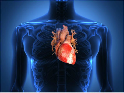
Echocardiography has become routinely used in the diagnosis, management, and follow-up of patients with any suspected or known heart diseases. It is one of the most widely used diagnostic tests in cardiology. It can provide a wealth of helpful information, including the size and shape of the heart, pumping capacity, and the location and extent of any tissue damage. An Echocardiogram can also give physicians other estimates of heart function such as a calculation of the cardiac output, ejection fraction, and diastolic function (how well the heart relaxes).
Echocardiography can help detect cardiac tumours, congenital malformation, cardiomyopathies, such as hypertrophic cardiomyopathy, dilated cardiomyopathy, and many others. The use of Stress Echocardiography may also help determine whether any chest pain or associated symptoms are related to heart disease. The biggest advantage to echocardiography is that it is non-invasive (doesn’t involve breaking the skin or entering body cavities) and has no known risks or side effects. Because it is an ultrasound, it does not give off any radiation.
Not only can an echocardiogram create ultrasound images of heart structures, but it can also produce accurate assessment of the blood flowing through the heart, using pulsed or continuous wave Doppler ultrasound. This allows assessment of both normal and abnormal blood flow through the heart. Color Doppler as well as spectral Doppler is used to visualize any abnormal communications between the left and right side of the heart, any leaking of blood through the valves (valvular regurgitation), and to estimate how well the valves open (or do not open in the case of valvular stenosis).
Echocardiography was also the first ultrasound subspecialty to use intravenous contrast. (See Contrast Echocardiography).
Echocardiography is performed by cardiac sonographers or doctors trained in echocardiography.
-
Why It Is Done
The Echocardiogram is done to:
· Look for the cause of abnormal heart sounds (murmurs or clicks), an enlarged heart, unexplained chest pains, shortness of breath, fainting spells or irregular heartbeats.
· Check the thickness and movement of the heart wall.
· Look at the heart valves and check how well they work.
· See how well an artificial heart valve is working.
· Measure the size and shape of the heart’s chambers.
· Check the ability of your heart chambers to pump blood (cardiac performance). During an echocardiogram, your doctor can calculate how much blood your heart is pumping during each heartbeat (ejection fraction). You might have a low ejection fraction if you have heart failure.
· Detect a disease that affects the heart muscle and the way it pumps, such as cardiomyopathy.
· Look for blood clots and tumours inside the heart.
· Look for congenital heart defects or to check the effectiveness of previous surgery to repair a congenital heart defect.
· Check how well your heart works after a heart attack.
· Identify the specific cause of heart failure.
· Look for a collection of fluid around the heart (pericardial effusion).
· Look for a thickening of the lining (pericardium) around the heart.Doppler echocardiogram can be done during an echocardiogram or a stress echocardiogram to:
· Measure the speed at which blood travels through the heart.
· Measure the blood pressure and speed of blood flow through the heart valves. -
How to prepare
You do not need any special preparation for an echocardiogram.
-
How it is done
An echocardiogram may be done in a hospital, clinic, or doctor’s office. It can also be done at your bedside in the hospital.
You will need to remove any jewellery and clothes above your waist (you may be allowed to keep on your underwear if it does not interfere with the test). You will be given a special gown appropriate to the test.
You will lie on your back or on your left side on a bed or table. Small electrodes will be put to your arms and torso to record your heart rate during the test. A small amount of gel will be rubbed on the left side of your chest to help pick up the sound waves. A small transducer is pressed firmly against your chest and moved slowly back and forth. The transducer sends sound waves into the chest and picks up the echoes as they reflect off different parts of the heart. The echoes are sent to a video monitor that records pictures of your heart for later viewing and evaluation. The room is usually darkened to help the technician see the pictures on the monitor.
At times you will be asked to hold very still, breathe in and out very slowly, hold your breath, or lie on your left side. The transducer is usually moved to different areas on your chest that provides specific views of your heart.
The test usually takes from 30 minutes. When the test is over, the gel is wiped off and the electrodes are removed.
-
How it feels
You will not have pain from the echocardiogram. Gel is put on your chest for the ultrasound. It may feel cool. The handheld ultrasound device is pressed firmly against your chest, but it does not cause pain. You will not hear or feel the sound waves.
Most people do not experience any discomfort from ultrasound tests. But if you have severe difficulty breathing or cannot lie flat for a long exam, you may not be able to have an entire echo study. Talk to your doctor or the technician performing your echo about any concerns you have.
-
Risks
An echocardiogram is safe, because the test uses only sound waves to evaluate your heart. These high-frequency sound waves have not been shown to have any harmful effects.
-
Results
Results are usually available within one day. If the test is done by a cardiologist, the results may be available immediately after the test.
-
What affects the test
You may not be able to have the test or the results may not be helpful if you are:
· Overweight
· Have a thick chest or large breasts
· Are unable to sit still
· Have lung disease, such as chronic obstructive pulmonary disease (COPD)In these situations, other heart tests may be done:
· Cardiac Blood Pool Scan
· Electrocardiogram
· Exercise Electrocardiogram
· Cardiac Catheterization
Stress Echocardiography
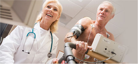 Stress echocardiogram, also known as a stress echo or SE, utilizes ultrasound imaging of the heart to assess the wall motion in response to physical stress. A stress echo is a non-invasive test and is performed in the presence of a licensed medical professional, such as a cardiologist, and a cardiac sonographer. First, images of the heart are taken “at rest” to acquire a baseline of the patient’s wall motion at a resting heart rate. The patient then walks on a treadmill or utilizes another exercise modality to increase the heart rate to his or her target heart rate, or 85% of the age predicted max heart rate. If the patient cannot exercise for any reason, he or she may be asked do consent to a chemically-induced stress test. Finally, images of the heart are taken “at stress” to assess wall motion at the peak heart rate. A stress echo assesses wall motion of the heart; it does not, however, image the coronary arteries directly. Ischemia of one or more coronary arteries could cause a wall motion abnormality which could indicate coronary artery disease (CAD). The gold standard test to directly image the coronary arteries and directly assess for stenosis or occlusion is a cardiac catheterization.
-
Why is it done
Stress echo may be done to:
· Identify and monitor reduced blood flow to heart muscle (ischemia). This is usually more apparent after some form of stress, such as exercise or special medicine.
· Evaluate diastolic dysfunction -
How to prepare
Stress echocardiogram
1. Do not eat heavily for a few hours before a stress echo to help prevent nausea. You may feel nauseated if you exercise with a full stomach or from the injection of dobutamine.
2. Wear flat, comfortable shoes (no bedroom slippers or sandals) and loose, lightweight shorts or sweatpants for an exercise stress echo.! Ask your doctor whether you should take your regular medicines as usual. Tell your doctor if you take insulin.
Before a stress echocardiogram, you will be asked to sign a consent form that says you understand the risks of the test and agree to have it done.
Talk to your doctor about any concerns you have regarding the need for the test, its risks, how it will be done, or what the results will mean. To help you understand the importance of this test, fill out the medical test information form.
-
How it is done
Stress echocardiogram may be done in a hospital, clinic, or doctor’s office. It can also be done at your bedside in the hospital.
You will need to remove any jewellery and clothes above your waist (you may be allowed to keep on your underwear if it does not interfere with the test). You will be given a special gown appropriate to the test.
Stress echocardiogram is usually performed by two trained ultrasound technicians. Prior to the procedure, the technician will explain how the test is going to be performed and will obtain your written consent. After that you will be connected to an ECG monitoring unit and will be asked to lie down on the bed. Blood pressure is monitored at times through the test. The resting ECG and resting echocardiogram views will be obtained. After that you will proceed to an exercising stage where you will run on the treadmill to reach the target heart rate. After it has been reached, you will return on to the bed and the technician will obtain stress echocardiography views. You will now proceed to recovery stage, at which time both technicians will make sure that you heart rate comes back to normal and will only finish the test after that.
In cases when your doctor determines that you cannot perform an exercise stress echocardiogram, he might refer you for a pharmaceutically (dobutamine or persantine) induced stress test. The difference between pharmaceutical and a conventional running test is that you don’t have to exercise; instead a special medicine is injected into the bloodstream by the means of an IV that will increase your heart rate to reach the targeted peak. The whole test is preceded with a patient lying down on a bed.
An exercise stress echo takes about 30 to 60 minutes.
-
How it feels
You will not have pain from the echocardiogram. Gel is put on your chest for the ultrasound. It may feel cool. The handheld ultrasound device is pressed firmly against your chest, but it does not cause pain. You will not hear or feel the sound waves. Most people do not experience any discomfort from ultrasound tests. But if you have severe difficulty breathing or cannot lie flat for a long exam, you may not be able to have an entire echo study.
If you are able to run on the treadmill, you will experience your heart racing and may experience shortness of breath, leg or knee pain, or chest pain.
Talk to your doctor or the technician performing your echo about any concerns you have.
-
Dobutamine/Persantine stress echocardiogram
· You may have a brief, sharp pain when the intravenous (IV) needle is placed in a vein in your arm.
· If medicine to stress your heart is used, you may have symptoms of mild nausea, headache, dizziness, flushing, or chest pain (angina). These symptoms only last a few minutes.
-
Risks
A stress echocardiogram can cause dizziness, low blood pressure, shortness of breath, nausea, irregular heartbeats.
-
Results
Results are usually available within one day. If the test is done by a cardiologist, the results may be available immediately after the test.
-
What affects the test
You may not be able to have the test or the results may not be helpful if you are:
· Overweight
· Have a thick chest or large breasts
· Are unable to sit still
· Have lung disease, such as chronic obstructive pulmonary disease (COPD).In these situations, other heart tests may be done:
· Cardiac Blood Pool Scan
· Electrocardiogram
· Exercise Electrocardiogram
· Cardiac Catheterization
· Transesophageal Echocardiogram
Three-Dimensional Echocardiography
 3D echocardiography (also known as 4D echocardiography when the picture is moving) is now possible, using a matrix array ultrasound probe and an appropriate processing system. This enables detailed anatomical assessment of cardiac pathology, particularly valvular defects, and cardiomyopathies. The ability to slice the virtual heart in infinite planes in an anatomically appropriate manner and to reconstruct three-dimensional images of anatomic structures makes 3D echocardiography unique for the understanding of the congenitally malformed heart. Real Time 3-Dimensional echocardiography can be used to guide the location of bioptomes during right ventricular endomyocardial biopsies, placement of catheter delivered valvular devices, and in many other intraoperative assessments.
-
Why it is done
The 3D Echocardiogram is done to:
· Look for the cause of abnormal heart sounds (murmurs or clicks), an enlarged heart, unexplained chest pains, shortness of breath, fainting spells or irregular heartbeats.
· Check the thickness and movement of the heart wall.
· Look at the heart valves and check how well they work.
· See how well an artificial heart valve is working.
· Measure the size and shape of the heart’s chambers.
· Check the ability of your heart chambers to pump blood (cardiac performance). During an echocardiogram, your doctor can calculate how much blood your heart is pumping during each heartbeat (ejection fraction). You might have a low ejection fraction if you have heart failure.
· Detect a disease that affects the heart muscle and the way it pumps, such as cardiomyopathy.
· Look for blood clots and tumours inside the heart.
· Look for congenital heart defects or to check the effectiveness of previous surgery to repair a congenital heart defect.
· Check how well your heart works after a heart attack.
· Identify the specific cause of heart failure.
· Look for a collection of fluid around the heart (pericardial effusion).
· Look for a thickening of the lining (pericardium) around the heart. -
How to prepare
You do not need any special preparation for a 3D echocardiogram.
-
How it is done
A 3D echocardiogram may be done in a hospital, clinic, or doctor’s office. It can also be done at your bedside in the hospital.
You will need to remove any jewellery and clothes above your waist (you may be allowed to keep on your underwear if it does not interfere with the test). You will be given a special gown appropriate to the test.
You will lie on your back or on your left side on a bed or table. Small electrodes will be put to your arms and legs to record your heart rate during the test. A small amount of gel will be rubbed on the left side of your chest to help pick up the sound waves. A small transducer is pressed firmly against your chest and moved slowly back and forth. The transducer sends sound waves into the chest and picks up the echoes as they reflect off different parts of the heart. The echoes are sent to a video monitor that records pictures of your heart for later viewing and evaluation. The room is usually darkened to help the technician see the pictures on the monitor.
At times you will be asked to hold very still, breathe in and out very slowly, hold your breath, or lie on your left side. The transducer is usually moved to different areas on your chest that provides specific views of your heart.
The test usually takes from 30 minutes. When the test is over, the gel is wiped off and the electrodes are removed.
-
How it feels
You will not have pain from the 3D echocardiogram. Gel is put on your chest for the ultrasound. It may feel cool. The handheld ultrasound device is pressed firmly against your chest, but it does not cause pain. You will not hear or feel the sound waves.
Most people do not experience any discomfort from ultrasound tests. But if you have severe difficulty breathing or cannot lie flat for a long exam, you may not be able to have an entire echo study. Talk to your doctor or the technician performing your echo about any concerns you have.
-
Results
Results are usually available within one day. If the test is done by a cardiologist, the results may be available immediately after the test.
-
What affects the test
You may not be able to have the test or the results may not be helpful if you are:
· Overweight
· have a thick chest or large breasts
· Are unable to sit still
· have lung disease, such as chronic obstructive pulmonary disease (COPD).In these situations, other heart tests may be done:
· Cardiac Blood Pool Scan
· Electrocardiogram
· Exercise Electrocardiogram
· Cardiac Catheterization
Contrast Echocardiography
 Contrast echocardiography, or Contrast-enhanced ultrasound is the addition of ultrasound contrast medium, or imaging agent, to traditional ultrasonography. The ultrasound contrast is made up of tiny microbubbles filled with a gas core and protein shell. This allows the microbubbles to circulate through the cardiovascular system and return the ultrasound waves creating a highly reflective image.
There are multiple applications in which contrast-enhanced ultrasound can be useful. The most commonly used application is in the enhancement of LV endocardial borders for assessment of global and regional systolic function. Contrast may also be used to enhance visualization of wall thickening during stress echocardiography, for the assessment of LV thrombus, or for the assessment of other masses in the heart. Contrast echocardiography has also been used to assess blood perfusion throughout myocardium in the case of coronary artery disease.
-
Risks
If contrast material is used, there is a slight risk of having an allergic reaction. Most reactions can be controlled using medicine.
-
Results
Results are usually available within one day. If the test is done by a cardiologist, the results may be available immediately after the test.
-
What affects the test
You may not be able to have the test or the results may not be helpful if you are:
· Overweight
· Have a thick chest or large breasts
· Are unable to sit still
· Have lung disease, such as chronic obstructive pulmonary disease (COPD).In these situations, other heart tests may be done:
· Cardiac Blood Pool Scan
· Electrocardiogram
· Exercise Electrocardiogram
· Cardiac Catheterization