What is ultrasound?
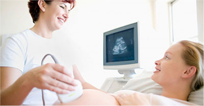 Ultrasound is safe and painless, and produces pictures of the inside of the body using sound waves. Ultrasound imaging, also called ultrasound scanning or sonography, involves the use of a small transducer (probe) and ultrasound gel placed directly on the skin. High-frequency sound waves are transmitted from the probe through the gel into the body. The transducer collects the sounds that bounce back and a computer then uses those sound waves to create an image. Ultrasound examinations do not use ionizing radiation (as used in x-rays), thus there is no radiation exposure to the patient. Because ultrasound images are captured in real-time, they can show the structure and movement of the body’s internal organs, as well as blood flowing through blood vessels.
Ultrasound imaging is a non-invasive medical test that helps physicians diagnose and treat medical conditions.
Conventional ultrasound displays the images in thin, flat sections of the body. Advancements in ultrasound technology include three-dimensional (3-D) ultrasound that formats the sound wave data into 3-D images.
A Doppler ultrasound study may be part of an ultrasound examination.
Doppler ultrasound is a special ultrasound technique that evaluates blood flow through a blood vessel, including the body’s major arteries and veins in the abdomen, arms, legs, neck and head (in infants and children).
There are three types of Doppler ultrasound:
• Color Doppleruses a computer to convert Doppler measurements into an array of colors to show the speed and direction of blood flow through a blood vessel.
• Power Doppleris a newer technique that is more sensitive than color Doppler and capable of providing greater detail of blood flow, especially when blood flow is little or minimal. Power Doppler, however, does not help the radiologist determine the direction of blood flow, which may be important in some situations.
• Spectral Dopplerdisplays blood flow measurements graphically, in terms of the distance traveled per unit of time, rather than as a color picture. It can also convert blood flow information into a distinctive sound that can be heard with every heartbeat.
Ultrasound is a useful way of examining many of the body’s internal organs, including but not limited to the:
• heart and blood vessels, including theabdominal aorta and its major branches
• liver
• gallbladder
• spleen
• pancreas
• kidneys
• bladder
• uterus,ovaries, and unborn child (fetus) in pregnant patients
• eyes
• thyroidand parathyroid glands
• scrotum(testicles)
• brain in infants
• hips in infants
Ultrasound is used to help physicians evaluate symptoms such as:
• pain
• swelling
• infection
Ultrasound is also used to:
• guide procedures such asneedle biopsies, in which needles are used to sample cells from an abnormal area for laboratory testing.
• image the breasts and guidebiopsy of breast cancer
• diagnose a variety of heart conditions, including valve problems and congestive heart failure, and to assess damage after a heart attack. Ultrasound of the heart is commonly called an “echocardiogram” or “echo”.
-
How should I prepare?
You should wear comfortable, loose-fitting clothing for your ultrasound exam. You may need to remove all clothing and jewelry in the area to be examined.
You may be asked to wear a gown during the procedure.Preparation for the procedure will depend on the type of examination you will have. For some scans your doctor may instruct you not to eat or drink for as many as 12 hours before your appointment. For others you may be asked to drink up to six glasses of water two hours prior to your exam and avoid urinating so that your bladder is full when the scan begins.
-
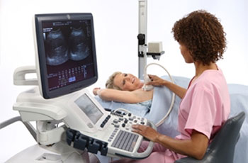
What does the equipment look like?
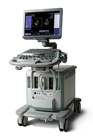
Ultrasound scanners consist of a console containing a computer and electronics, a video display screen and a transducer that is used to do the scanning. The transducer is a small hand-held device that resembles a microphone, attached to the scanner by a cord. The transducer sends out inaudible high frequency sound waves into the body and then listens for the returning echoes from the tissues in the body. The principles are similar to sonar used by boats and submarines.
The ultrasound image is immediately visible on a video display screen that looks like a computer monitor. The image is created based on the amplitude (loudness), frequency (pitch) and time it takes for the ultrasound signal to return from the area of the patient being examined to the transducer (the device used to examine the patient), as well as the type of body structure and composition of body tissue through which the sound travels. A small amount of gel is put on the skin to allow the sound waves to travel back and forth from the transducer.
-
How does the procedure work?
Ultrasound imaging is based on the same principles involved in the sonar used by submarines. When a sound wave strikes an object, it bounces back, or echoes. By measuring these echo waves, it is possible to determine how far away the object is as well as the object’s size, shape and consistency (whether the object is solid or filled with fluid).
In medicine, ultrasound is used to detect changes in appearance, size or contour of organs, tissues, and vessels or detect abnormal masses, such as tumors.
In an ultrasound examination, a transducer both sends the sound waves and receives the echoing waves. When the transducer is pressed against the skin, it directs small pulses of inaudible, high-frequency sound waves into the body. As the sound waves bounce off internal organs, fluids and tissues, the sensitive microphone in the transducer records tiny changes in the sound’s pitch and direction. These signature waves are instantly measured and displayed by a computer, which in turn creates a real-time picture on the monitor. One or more frames of the moving pictures are typically captured as still images. Small loops of the moving “real time” images may also be saved.
Doppler ultrasound, a special application of ultrasound, measures the direction and speed of blood cells as they move through vessels. The movement of blood cells causes a change in pitch of the reflected sound waves (called the Doppler effect). A computer collects and processes the sounds and creates graphs or color pictures that represent the flow of blood through the blood vessels.
-
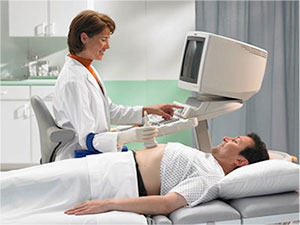
How is the procedure performed?
For most ultrasound exams, you will be positioned lying face-up on an examination table that can be tilted or moved.
After you are positioned on the examination table, the sonographer will apply a warm water-based gel to the area of the body being studied. The gel will help the transducer make secure contact with the body and eliminate air pockets between the transducer and the skin that can block the sound waves from passing into your body. The transducer is placed on the body and moved back and forth over the area of interest until the desired images are captured.
There is usually no discomfort from pressure as the transducer is pressed against the area being examined. However, if scanning is performed over an area of tenderness, you may feel pressure or minor pain from the transducer.
Doppler sonography is performed using the same transducer.
In some ultrasound studies, the transducer is attached to a probe and inserted into a natural opening in the body. These exams include:
• Transrectal ultrasound: The transducer is inserted into a man’s rectum to view the prostate.
• Transvaginal ultrasound: The transducer is inserted into a woman’s vagina to view the uterus and ovaries.
-
What will I experience during and after the procedure?
Ultrasound examinations are painless and easily tolerated by most patients.
Ultrasound exams in which the transducer is inserted into an opening of the body may produce minimal discomfort.
Most ultrasound examinations are completed within 30 minutes, although more extensive exams may take up to an hour.
When the examination is complete, you may be asked to dress and wait while the ultrasound images are reviewed.
After an ultrasound examination, you should be able to resume your normal activities immediately.
-
Who interprets the results and how do I get them?
A radiologist, a physician specifically trained to supervise and interpret radiology examinations, will analyze the images and send a signed report to your primary care physician, or to the physician or other healthcare provider who requested the exam, and he/she will share the results with you. In some cases the radiologist may discuss results with you at the conclusion of your examination.
Follow-up examinations may be necessary, and your doctor will explain the exact reason why another exam is requested. Sometimes a follow-up exam is done because a suspicious or questionable finding needs clarification with additional views or a special imaging technique. A follow-up examination may also be necessary so that any change in a known abnormality can be monitored over time. Follow-up examinations are sometimes the best way to see if treatment is working or if an abnormality is stable over time.
-
What are the benefits vs. risks?
Benefits
• Most ultrasound procedures are non-invasive (no needles or injections).
• Occasionally, an ultrasound exam may be temporarily uncomfortable, but it is almost never painful.
• Ultrasound is widely available, easy-to-use and less expensive than other imaging methods.
• Ultrasound imaging is extremely safe and does not use any ionizing radiation.
• Ultrasound scanning gives a clear picture of soft tissues that do not show up well on x-ray images.
• Ultrasound is the preferred imagingmodality for the diagnosis and monitoring of pregnant women and their unborn babies.
• Ultrasound provides real-time imaging, making it a good tool for guidingminimally invasive procedures such as needle biopsies and fluid aspiration.
Risks
• For standarddiagnostic ultrasound, there are no known harmful effects on humans.
-
What are the limitations of Ultrasound Imaging?
Ultrasound waves are disrupted by air or gas; therefore ultrasound is not an ideal imaging technique for air-filled bowel or organs obscured by the bowel. In most cases, barium exams, CT scanning, and MRI are the methods of choice in such a setting.
Large patients are more difficult to image by ultrasound because greater amounts of tissue attenuates (weakens) the sound waves as they pass deeper into the body.
Ultrasound has difficulty penetrating bone and, therefore, can only see the outer surface of bony structures and not what lies within (except in infants who have more cartilage in their skeletons than older children or adults). For visualizing internal structure of bones or certain joints, other imaging modalities such as MRI are typically used.
Abdomen Ultrasound
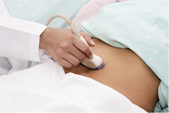
 An abdominal ultrasound produces a picture of the organs and other structures in the upper abdomen.
Abdominal ultrasound imaging is performed to evaluate:
• kidneys
• liver
• gallbladder
• pancreas
• spleen
• abdominal aorta and other blood vessels of the abdomen
-
Uses
Ultrasound is used to:
• Find the possible cause of abdominal pain.
• Find, measure, or monitor an aneurysm in the aorta. An aneurysm may cause a large, pulsing lump in the abdomen.
• Check the size, shape, and position of the An ultrasound may be done to evaluate jaundice and other problems of the liver, including liver masses, cirrhosis, fat deposits in the liver (called fatty liver), or abnormal liver function tests.
• Detect gallstones, inflammation of the gallbladder (cholecystitis), or blocked bile ducts.
• Learn the size of an enlarged spleen and look for damage or disease.
• Find problems with the pancreas, such as a pancreatic tumor.
• Look for blocked urine flow in a A kidney ultrasound may also be done to find out the size of the kidneys, detect kidney masses, detect fluid surrounding the kidneys, investigate causes for recurring urinary tract infections, or check the condition of transplanted kidneys.
• Find out whether a mass in any of the abdominal organs (such as the liver) is a solid tumor or a simple fluid-filled
• Guide the placement of a needle or other instrument during biopsy.
• Look for fluid buildup in the abdominal cavity (ascites). An ultrasound also may be done to guide the needle during a procedure to remove fluid from the abdominal cavity (paracentesis).
-
How should I prepare?
Tell your doctor if you have had a barium enema or a series of upper GI (gastrointestinal) tests within the past 2 days. Barium that remains in the intestines can interfere with the ultrasound test.
Preparations depend on the type of ultrasound you are having.
• For abdominal ultrasound, you will need to avoid eating for eight to 12 hours before the test.
• If the doctor has requested Abdomen and Pelvic ultrasound, then in addition to stop eating 8 to 12 hours before the test, you will be asked to drink 6-8 glasses of liquid one hour prior to the exam and not void.
-
How the test is done
This test is done by an ultrasound technologist (sonographer). It is done in an ultrasound room.
You will need to take off any jewelry that might interfere with the ultrasound scan. You will need to take off some of your clothes, depending on which area is examined (you may be allowed to keep on your underwear if it does not interfere with the test). You will be given a cloth or paper covering to use during the test.
During the test, you will lie on your back (or on your side) on a padded exam table. Warmed gel will be spread on your abdomen. A small handheld unit called a transducer is pressed against your abdomen.
You need to lie very still while the ultrasound scan is being done. You may be asked to take a breath and hold it for several seconds during the scanning. This lets the sonographer to see organs and structures, such as the bile ducts, more clearly because they are not moving. Holding your breath also temporarily pushes the liver and spleen lower into the belly so they are not hidden by the lower ribs, which makes it harder for the sonographer to see them clearly.
Abdominal ultrasound usually takes 30 to 60 minutes.
Arterial Ultrasound
 Ultrasound is safe and painless, and produces pictures of the inside of the body using sound waves. Ultrasound imaging is a non-invasive medical test that helps physicians diagnose and treat medical conditions.
Arterial ultrasound provides pictures of the veins throughout the body. A Doppler ultrasound study may be part of an arterial ultrasound examination. Doppler ultrasound is a special ultrasound technique that evaluates blood flow through a blood vessel, including the body’s major arteries and veins in the abdomen, arms, legs, neck and head (in infants and children).
The purpose of a lower and upper extremity arterial evaluation is to detect the presence, severity and location of atherosclerosis (narrowing of the arteries caused by plaque) in your legs and arms. Some of the indications for an arterial lower extremity evaluation include leg pain while walking (claudication), leg pain at rest, leg numbness and tingling, or non-healing ulcers or sores of the legs or feet. Upper extremity arterial ultrasound indications include but are not limited to: diabetes, numbness, upper limbs pain.
-
How should I prepare?
You should wear comfortable, loose-fitting clothing for your ultrasound exam. You may need to remove all clothing and jewellery in the area to be examined. You may be asked to wear a gown during the procedure.
-
How is the procedure performed?
For most ultrasound exams, you will be positioned lying face-up on an examination table that can be tilted or moved. A clear water-based gel is applied to the area of the body being studied to help the transducer make secure contact with the body and eliminate air pockets between the transducer and the skin that can block the sound waves from passing into your body. The sonographer (ultrasound technologist) or radiologist then places the transducer on the skin in various locations, sweeping over the area of interest or angling the sound beam from a different location to better see an area of concern.
Doppler sonography is performed using the same transducer.
When the examination is complete, you may be asked to dress and wait while the ultrasound images are reviewed.
This ultrasound examination is usually completed within 30 to 45 minutes. More complex exams may take a longer period of time.
If a Doppler ultrasound study is performed, you may actually hear pulse-like sounds that change in pitch as the blood flow is monitored and measured.
Once the imaging is complete, the clear ultrasound gel will be wiped off your skin. Any portions that are not wiped off will dry to a powder. The ultrasound gel does not stain or discolour clothing.
-
Who interprets the results and how do I get them?
A radiologist, a physician specifically trained to supervise and interpret radiology examinations, will analyze the images and send a signed report to your primary care physician, or to the physician or other healthcare provider who requested the exam, and he/she will share the results with you.
Follow-up examinations may be necessary, and your doctor will explain the exact reason why another exam is requested. Sometimes a follow-up exam is done because a suspicious or questionable finding needs clarification with additional views or a special imaging technique. A follow-up examination may also be necessary so that any change in a known abnormality can be monitored over time. Follow-up examinations are sometimes the best way to see if treatment is working or if an abnormality is stable over time.
Breast Ultrasound
 Ultrasound imaging of the breast produces a picture of the internal structures of the breast.
During a breast ultrasound examination the sonographer or physician performing the test may use Doppler techniques to evaluate blood flow or lack of flow in any breast mass. In some cases this may provide additional information as to the cause of the mass.
-
What are some common uses of the procedure?
• Determining the Nature of a Breast Abnormality
The primary use of breast ultrasound today is to help diagnose breast abnormalities detected by a physician during a physical exam (such as a lump or bloody or spontaneous clear nipple discharge) and to characterize potential abnormalities seen on mammography or breast magnetic resonance imaging (MRI).
Ultrasound imaging can help to determine if an abnormality is solid (which may be a non-cancerous lump of tissue or a cancerous tumor) or fluid-filled (such as a benign cyst) or both cystic and solid. Ultrasound can also help show additional features of the abnormal area.
Doppler ultrasound is used to assess blood supply in breast lesions.
• Supplemental Breast Cancer Screening
Mammography and 3D Breast Ultrasound are known to reduce deaths due to breast cancer through early detection. Even so, mammograms do not detect all breast cancers. Some breast lesions and abnormalities are not visible or are difficult to interpret on mammograms. In breasts that are dense, meaning there is a lot of ducts, glands, fibrous tissue and less fat, many cancers can be hard to see on mammography. In this case, 3D Breast Ultrasound is recommended as it is proven to provide highly accurate diagnostic information for patients with dense or small breast.
• For more information on 3D Breast Screening, please visit: canadianbreastscreening.com/new-safe-choice/
• Ultrasound-guided Breast Biopsy
When an ultrasound examination reveals a suspicious breast abnormality, a physician may choose to perform an ultrasound-guided biopsy. Because ultrasound provides real-time images, it is often used to guide biopsy procedures. An ultrasound exam will usually need to be performed before the biopsy in order to plan the procedure and to determine if this method of biopsy can be used.
-
How should I prepare?
You will be asked to undress from the waist up and to wear a gown during the procedure. No other special preparation is needed.
-
How is the procedure performed?
You will lie on your back on the examining table and may be asked to raise your arm above your head.
After you are positioned on the examination table, the radiologist or sonographer will apply a warm water-based gel to the area of the body being studied. The gel will help the transducer make secure contact with the body and eliminate air pockets between the transducer and the skin that can block the sound waves from passing into your body. The transducer is placed on the body and moved back and forth over the area of interest until the desired images are captured.
There is usually no discomfort from pressure as the transducer is pressed against the area being examined. However, if scanning is performed over an area of tenderness, you may feel pressure or minor pain from the transducer.
Once the imaging is complete, the clear ultrasound gel will be wiped off your skin. Any portions that are not wiped off will dry to a powder. The ultrasound gel does not stain or discolor clothing.
-
How long does a procedure take?
The conventional (2D) ultrasound usually takes about 35-30 minutes for both breasts. A 3D procedure takes about 20 minutes.
Carotid Ultrasound
 Human’s body has two carotid arteries, which are located on the each side of the neck and carry blood from the heart to the brain. Ultrasound provides detailed pictures of these blood vessels and information about the blood flowing through them.
A Doppler ultrasound study is usually an integral part of a carotid ultrasound examination.
Doppler ultrasound is a special ultrasound technique that evaluates blood flow through the blood vessel, including the body’s major arteries and veins in the abdomen, arms, legs, neck and head (in infants and children).
-
What are some common uses of the procedure?
The carotid ultrasound is most frequently performed to detect narrowing, or stenosis, of the carotid artery, a condition that substantially increases the risk of stroke.
The major goal of carotid ultrasound is to screen patients for blockage or narrowing of their carotid arteries, which, if present, may increase their risk of having a stroke. If a significant narrowing is detected, a comprehensive treatment may be initiated.
It may also be performed if a patient has high blood pressure or a carotid bruit – an abnormal sound in the neck that is heard with the stethoscope. In some cases, it is also performed in preparation for coronary artery bypass surgery.
Other risk factors calling for a carotid ultrasound are:
• diabetes
• elevated blood cholesterol
• a family history of stroke or heart disease
A carotid ultrasound is also performed to:
• locate a hematoma, a collection of clotted blood that may slow and eventually stop the blood flow
• check the state of the carotid artery after surgery to restore normal blood flow
• verify the position of a metal stent placed to maintain carotid blood flow
Doppler ultrasound images can help the physician to see and evaluate:
• blockages to blood flow (such as clots)
• narrowing of vessels
• tumours and congenital vascular malformations
-
How should I prepare?
You may need to remove all clothing and jewellery in the area to be examined. A loose-fitting, open necked shirt or blouse is ideal. No other preparation is required.
-
How is the procedure performed?
For most ultrasound exams, you will be positioned lying face-up on an examination table that can be tilted or moved.
A clear water-based gel is applied to the area of the body being studied to help the transducer make secure contact with the body and eliminate air pockets between the transducer and the skin that can block the sound waves from passing into your body. The sonographer (ultrasound technologist) or radiologist then places the transducer on the skin in various locations, sweeping over the area of interest or angling the sound beam from a different location to better see an area of concern.
Doppler sonography is performed using the same transducer.
When the examination is complete, you may be asked to dress and wait while the ultrasound images are reviewed.
This ultrasound examination is usually completed within 30 minutes.
-
Who interprets the results and how do I get them?
A radiologist, a physician specifically trained to supervise and interpret radiology examinations, will analyze the images and send a signed report to your primary care physician, or to the physician or other healthcare provider who requested the exam, and he/she will share the results with you.
Follow-up examinations may be necessary in some cases, and your doctor will explain the exact reason why another exam is requested. Sometimes a follow-up exam is done because a suspicious or questionable finding needs clarification with additional views or a special imaging technique. A follow-up examination may also be necessary so that any change in a known abnormality can be monitored over time. Follow-up examinations are sometimes the best way to see if treatment is working or if an abnormality is stable over time.
-
What are the benefits vs. risks?
Benefits
• Most ultrasound scanning is non-invasive (no needles or injections).
• Occasionally, an ultrasound exam may be temporarily uncomfortable, but it is almost never painful.
• Ultrasound is widely available, easy-to-use and less expensive than other imaging methods.
• Ultrasound imaging is extremely safe and does not use any ionizing radiation.
• Ultrasound scanning gives a clear picture of soft tissues that do not show up well on x-ray images.
• If a carotid ultrasound exam shows narrowing of one or both carotid arteries, treatment can be taken to restore the free flow of blood to the brain. Many strokes are prevented as a result.
Risks
For standard diagnostic ultrasound, there are no known harmful effects on humans. In nearly 50 years of experience, carotid ultrasound has proved to be a risk-free procedure.
-
What are the limitations of Carotid Ultrasound Imaging?
Carotid ultrasound may be difficult or impossible if a patient has a bandage covering a wound or surgical scar in the neck.
An occasional patient is difficult to examine because of the size or contour of the neck.
Calcium deposits in the wall of the carotid artery may make it difficult to evaluate the vessel.
A small amount of soft plaque that produces low-level echoes may go undetected.
Ultrasound cannot visualize the entire length of the vessel because the last portion of the carotid artery travels though the bone at the base of the skull. For a more complete assessment, patients may need to undergo a CT or MRI of the carotid arteries.
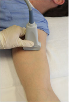
Musculoskeletal Ultrasound
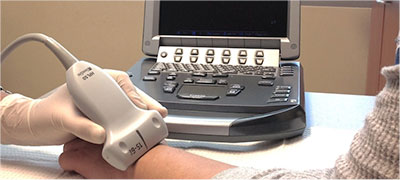
Ultrasound images of the musculoskeletal system provide pictures of muscles, tendons, ligaments, joints and soft tissue throughout the body.
-
What are some common uses of the procedure?
Ultrasound images are typically used to help diagnose:
• tendon tears, or tendinitis of the rotator cuff in the shoulder, Achilles tendon in the ankle and other tendons throughout the body.
• muscle tears, masses or fluid collections.
• ligament sprains or tears.
• inflammation or fluid (effusions) within the bursae and joints.
• early changes of rheumatoid arthritis.
• nerve entrapments such as carpal tunnel syndrome.
• benign and malignant soft tissue tumors.
• ganglion cysts.
• hernias
• foreign bodies in the soft tissues (such as splinters or glass).
-
How should I prepare?
You should wear comfortable, loose-fitting clothing for your ultrasound exam. You may need to remove all clothing and jewelry in the area to be examined.
You may be asked to wear a gown during the procedure.
-
What does the equipment look like?
Ultrasound scanners consist of a computer and a transducer that is used to do the scanning. The transducer is a small hand-held device that resembles a microphone, attached to the scanner by a cord. The transducer sends out inaudible high frequency sound waves into the body and then listens for the returning echoes from the tissues in the body. The principles are similar to sonar used by boats and submarines.
The ultrasound image is immediately visible on a video display screen. The image is created based on the amplitude (loudness), frequency (pitch) and time it takes for the ultrasound signal to return from the area of the patient being examined to the transducer (the device used to examine the patient), as well as the type of body structure and composition of body tissue through which the sound travels. A small amount of gel is put on the skin to allow the sound waves to travel back and forth from the transducer.
-
How is the procedure performed?
For certain ultrasound examinations of the musculoskeletal system, the patient may be seated on an examination table or a swivel chair. For other ultrasound exams, the patient is positioned lying face-up or face-down on an examination table. The radiologist or sonographer may ask you to move the extremity being examined or may move it for you to evaluate the anatomy and function of the joint, muscle, ligament or tendon.
-
What will I experience during and after the procedure?
Ultrasound examinations are painless and easily tolerated by most patients.
Musculoskeletal ultrasound examination is usually completed within 15 to 30 minutes but may occasionally take longer.
When the examination is complete, you may be asked to dress and wait while the ultrasound images are reviewed.
After an ultrasound examination, you should be able to resume your normal activities immediately.
-
What are the limitations of Ultrasound Imaging of the Musculoskeletal System?
Ultrasound has difficulty penetrating bone and, therefore, can only see the outer surface of bony structures and not what lies within (except in infants who have more cartilage in their skeletons than older children or adults). For visualizing internal structure of bones or certain joints, other imaging modalities such as MRI are typically used.
There are also limitations to the depth that sound waves can penetrate; therefore, deeper structures in larger patients may not be seen easily.
Ultrasound has not proven useful in detecting whiplash injuries or most other causes of back pain.
Obstetric Ultrasound
 Obstetrical ultrasound provides pictures of an embryo or fetus within a woman’s uterus, as well as the mother’s uterus and ovaries.
During an obstetrical ultrasound the examiner may evaluate blood flow in the umbilical cord or may, in some cases, assess blood flow in the fetus or placenta.
-
What are some common uses of the procedure?
Obstetrical ultrasound is a useful clinical test to:
• establish the presence of a living embryo/fetus
• estimate the age of the pregnancy
• diagnose congenitalabnormalities of the fetus
• evaluate the position of the fetus
• evaluate the position of the placenta
• determine if there are multiple pregnancies
• determine the amount of amniotic fluid around the baby
• check for opening or shortening of the cervix
• assess fetal growth
• assess fetal well-being
Different information is gained at different times (trimesters) during the pregnancy.
• 1st-trimester fetal ultrasound is done to:
· Determine how pregnancy is progressing.
· Find out if there is more than 1 fetus.
· Estimate the age of the fetus (gestational age).
· Estimate the risk of a chromosomedefect, such as Down syndrome.
· Check for birth defects that affect the brain or spinal cord.
• 2nd-trimester fetal ultrasound is done to:
· Estimate the age of the fetus (gestational age).
· Look at the size and position of the fetus, placenta, and amniotic fluid.
· Determine the position of the fetus, umbilical cord, and the placenta during a procedure, such as an amniocentesisor umbilical cord blood sampling.
· Detect major birth defects, such as a neural tube defect or heart problems.
• 3rd-trimester fetal ultrasound is done to:
· Make sure that a fetus is alive and moving.
· Look at the size and position of the fetus, placenta, and amniotic fluid.
-
How should I prepare?
You should wear a loose-fitting, two-piece outfit for the examination. Only the lower abdominal area needs to be exposed during this procedure.
Sometimes the radiologist determines that a transvaginal scan needs to be performed in order to see the pregnancy more closely or to assess the cervix. This technique often provides improved, more detailed images of the uterus and ovaries. It is especially useful in early pregnancy.
For more information on transvaginal ultrasound, please see the Pelvic Ultrasound section.
-
How is the procedure performed?
You will be positioned lying face-up on an examination table that can be tilted or moved.
After you are positioned on the examination table, the sonographer will apply a warm water-based gel to the lower abdomen area. The gel will help the transducer make secure contact with the body and eliminate air pockets between the transducer and the skin that can block the sound waves from passing into your body. The transducer is placed on the body and moved back and forth over the area of interest until the desired images are captured.
There is usually no discomfort from pressure as the transducer is pressed against the area being examined.
-
What will I experience during and after the procedure?
Ultrasound examinations are painless and easily tolerated by most patients.
However, at times during an obstetrical ultrasound, the sonographer may have to press more firmly to get closer to the embryo or fetus to visualize the structure better. Any discomfort is usually minimal and temporary.
With transvaginal scanning, there may be minimal discomfort as the transducer is inserted into the vagina.
Obstetric ultrasound examination is usually completed within 30 minutes.
When the examination is complete, you may be asked to dress and wait while the ultrasound images are reviewed.
After an ultrasound examination, you should be able to resume your normal activities immediately.
-
What are the limitations of Obstetrical Ultrasound Imaging?
Obstetric ultrasound cannot identify all fetal abnormalities. Consequently, when there are clinical or laboratory suspicions for a possible abnormality, a pregnant woman may have to undergo non-radiologic testing such as amniocentesis (the evaluation of fluid taken from the sac surrounding the fetus) or chorionic villus sampling (evaluation of placental tissue) to determine the health of the fetus, or she may be referred by her primary care provider to a perinatologist (an obstetrician specializing in high-risk pregnancies).
Nuchal Translucency
The nuchal translucency screening is a test done during pregnancy. It uses ultrasound to measure the thickness of the fluid buildup at the back of the developing baby’s neck. If this area is thicker than normal, it can be an early sign of Down syndrome, trisomy 18, or heart problems.
The test is done between 11 and 14 weeks of pregnancy. It may be done as part of the first trimester screening test or the integrated screening test.
This test shows the chance that a baby may have a certain problem. Usually chorionic villus sampling (CVS) or amniocentesis is used to confirm if the baby actually has a problem.
-
Why it is done
A nuchal translucency test is done to find out the chance that your developing baby (fetus) may be at risk for having Down syndrome or other problems.
-
How to prepare
You may need a full bladder for the nuchal translucency test. If so, you’ll be asked to drink water or other liquids just before the test and to avoid urinating before the test.
Talk with your doctor about any concerns you have regarding the need for the test, its risks, how it will be done, or what the results will mean.
-
How it is done
Most often, a nuchal translucency test is done by a specially trained ultrasound technologist. But it may be done by a radiologist or an obstetrician who has received special training to do this test.
Often you don’t need to remove your clothes for the test. You can lift your shirt and push down the waistband of your skirt or pants. If you’re wearing a dress, you’ll be given a cloth or paper covering to put over your legs during the test.
During the test:
• You’ll lie down on your back or on your side on an exam table.
• A gel will be spread on your belly.
• A small, handheld device called a transducer will be pressed against the gel on your skin and moved over your belly. Images of the baby are displayed on a monitor. The technologist or doctor will look for and measure the thickness of the fluid buildup at the back of the baby’s neck.
When the test is done, the gel is wiped off your skin. You can urinate as soon as the test is done.
The test usually takes about 15 to 20 minutes.
-
How it feels
During a nuchal translucency test, you may have a feeling of pressure in your bladder. The gel may feel cool when it is first put on your belly. You’ll feel a light pressure from the transducer as it passes over your belly.
-
Risks
There are no known risks linked with a nuchal translucency test, either to you or the baby. But you may feel anxious if the test shows there is an increased chance that your baby may have a problem.
-
Results
Your doctor will look at the results of the nuchal translucency test to see if the area at the back of the baby’s neck is thicker than normal.
Normal: 2.5 millimeters (mm) or less
Abnormal: More than 2.5 mm
You may not receive information about the test right away. Full results are usually available in 1 or 2 days.
-
How accurate is the test?
The nuchal translucency test can find out if your developing baby (fetus) is at higher-than-normal risk for problems. But it can’t tell for sure that the baby has a problem. You would need to undergo another test for confirmation.
The accuracy of this test is based on how often the test correctly finds a problem. For example:
• The nuchal translucency test correctly finds Down syndrome in 64 to 70 out of 100 fetuses who have it. It misses Down syndrome in 30 to 36 out of 100 fetuses.
• First-trimester screening (nuchal translucency combined with bloodtests) correctly finds Down syndrome in 82 to 87 out of 100 fetuses who have it. But these tests miss it in 13 to 18 out of 100 fetuses.
• The integrated screening test(first-trimester tests plus the triple or quad screening blood tests in the second trimester) correctly finds Down syndrome in about 95 out of 100 fetuses who have it. This means that the test misses Down syndrome in 5 out of 100 fetuses.
Pelvis Ultrasound
 Ultrasound is safe and painless non-invasive procedure that produces pictures of the inside of the body using sound waves. Ultrasound examinations do not use ionizing radiation (as used in x-rays), thus there is no radiation exposure to the patient. Because ultrasound images are captured in real-time, they can show the structure and movement of the body’s internal organs, as well as blood flowing through blood vessels.
A pelvic ultrasound provides pictures of the structures and organs in the lower abdomen and pelvis.
There are three types of pelvic ultrasound:
• abdominal (transabdominal)
• vaginal (transvaginal, endovaginal) for women
• rectal (transrectal) for men
-
What are some common uses of the procedure?
In women, a pelvic ultrasound is most often performed to evaluate the:
• uterus
• cervix
• ovaries
• fallopian tubes
• bladderPelvic ultrasound exams are also used to monitor the health and development of an embryo or fetus during pregnancy. See the Obstetrical Ultrasound for more information.
Ultrasound examinations can help diagnose symptoms experienced by women such as:
• pelvic pain
• abnormal bleeding
• other menstrual problemsand help identify:
• palpablemasses such as ovarian cysts and uterine fibroids
• ovarian or uterinecancersA transvaginal ultrasound is usually performed to view the endometrium, the lining of the uterus, and the ovaries. Transvaginal ultrasound also provides a good way to evaluate the muscular walls of the uterus, called the myometrium. Sonohysterography allows for a more in-depth investigation of the uterine cavity. Three-dimensional (3D) ultrasound permits evaluation of the uterus and ovaries in planes that cannot be imaged directly. These exams are typically performed to detect:
• uterine anomalies
• uterine scars
• endometrial polyps
• fibroids
• cancer, especially in patients with abnormal uterine bleedingSome physicians also use 3D ultrasound or sonohysterography for patients with infertility. Three-dimensional ultrasound provides information about the outer contour of the uterus and about uterine irregularities.
In men, a pelvic ultrasound is used to evaluate the:
• bladder
• seminal vesicles
• prostateTransrectal ultrasound, a special study usually done to view the prostate gland, involves inserting a specialized ultrasound transducer into a man’s rectum. See Prostate Ultrasound for more information.
In men and women, a pelvic ultrasound exam can help identify:
• kidneystones
• bladdertumors
• other disorders of the urinary bladderDoppler ultrasound images can help the physician to see and evaluate:
• blockages to blood flow (such as clots).
• narrowing of vessels.
• tumors and congenital vascular malformations. -
How should I prepare?
For a pelvic ultrasound you need to drink at least 6 glasses of water within one hour prior to your examination and not void. You will be able to void in the middle of the exam. You might want to wear comfortable, loose-fitting clothing for your ultrasound exam. You may need to remove all clothing and jewelry in the area to be examined.
You may be asked to wear a gown during the procedure.
-
How is the procedure performed?
For women, the ultrasound is usually done with transabdominal probe first. The technician will scan with a probe for about 10-15 minutes, after which time you will be allowed to void and return for the following endovaginal scan with a different transducer. After this part is finished, your test is complete and technician will inform that you may now dress and are free to go.
For men, the ultrasound is usually done transabdominal, but in some cases in order to obtain better views, the transrectal scan is required.
The detailed description of these techniques are discussed as follows.
Transabdominal:
You will be positioned lying face-up on an examination table that can be tilted or moved.
After you are positioned on the examination table, the sonographer will apply a warm water-based gel to the area of the body being studied. The gel will help the transducer make secure contact with the body and eliminate air pockets between the transducer and the skin that can block the sound waves from passing into your body. The transducer is placed on the body and moved back and forth over the area of interest until the desired images are captured.
There is usually no discomfort from pressure as the transducer is pressed against the area being examined. However, if scanning is performed over an area of tenderness, you may feel pressure or minor pain from the transducer.
Once the imaging is complete, the clear ultrasound gel will be wiped off your skin. Any portions that are not wiped off will dry to a powder. The ultrasound gel does not stain or discolor clothing.
Transvaginal:
Transvaginal ultrasound is performed very much like a gynecologic exam and involves the insertion of the transducer into the vagina after you empty your bladder. The tip of the transducer is smaller than the standard speculum used when performing a Pap test. A protective cover is placed over the transducer, lubricated with a small amount of gel, and then inserted into the vagina. Only two to three inches of the transducer end are inserted into the vagina. The images are obtained from different orientations to get the best views of the uterus and ovaries. Transvaginal ultrasound is usually performed with you lying on your back, possibly with your feet in stirrups similar to a gynecologic exam.
Transrectal:
For a transrectal ultrasound, a protective cover is placed over the transducer. It is lubricated and then placed into the rectum.
Usually, you lie on your side, facing away from the examiner, with your knees and hips slightly flexed.
Doppler sonography is performed using the same transducer.
When the examination is complete, you may be asked to dress and wait while the ultrasound images are reviewed.
-
What will I experience during and after the procedure?
For a transabdominal exam:
A clear water-based gel is applied to the lower abdomen. A transducer is moved back and forth over this area until the desired images are captured.
Transabdomen examinations are painless and easily tolerated by most patients.
For a transvaginal exam:
With transvaginal ultrasound, although the examination is often performed to look for a cause of pelvic pain, the sonogram itself should not be painful or significantly increase your discomfort. A vaginal sonogram is usually more tolerable than a manual gynecologic examination.
For a transrectal exam:
If no biopsy is required, transrectal ultrasound of the prostate is similar to or may have less discomfort than a rectal exam performed by your doctor.
If a biopsy is performed, additional discomfort, due to the needle insertion, is usually minimal because the rectal wall is relatively insensitive to the pain in the region of the prostate. A biopsy will add time to the procedure.
If a Doppler ultrasound study is performed, you may actually hear pulse-like sounds that change in pitch as the blood flow is monitored and measured.
These ultrasound examinations are usually completed within 30 minutes.
After an ultrasound examination, you should be able to resume your normal activities immediately.
-
What are the benefits vs. risks?
Benefits
• Most ultrasound scanning is non-invasive (no needles or injections).
• Occasionally, an ultrasound exam may be temporarily uncomfortable, but it is almost never painful.
• Ultrasound is widely available, easy-to-use and less expensive than other imaging methods.
• Ultrasound imaging is extremely safe and does not use any ionizing radiation.
• Ultrasound scanning gives a clear picture of soft tissues that do not show up well on x-ray images.
• Ultrasound is the preferred imaging modality for the diagnosis and monitoring of pregnant women and their unborn babies.
• Ultrasound provides real-time imaging, making it a good tool for guiding minimally invasive procedures such as needle biopsies and fluid aspiration.
• Pelvic ultrasound can help to identify and evaluate a variety of urinary and reproductive system disorders in both sexes without even the minimal risks associated with x-ray
Risks
• For standard diagnostic ultrasound, there are no known harmful effects on humans.
-
What are the limitations of Pelvic Ultrasound Imaging?
Ultrasound waves are disrupted by air or gas; therefore ultrasound is not an ideal imaging technique for air-filled bowel or organs obscured by the bowel. In most cases, barium exams, CT scanning, and MRI are the methods of choice in such a setting.
Large patients are more difficult to image by ultrasound because greater amounts of tissue attenuates (weakens) the sound waves as they pass deeper into the body.
Scrotum Ultrasound
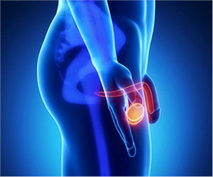 Ultrasound imaging of the scrotum provides pictures of the testicles and the surrounding tissues of a man or a boy. Ultrasound examinations do not use ionizing radiation (as used in x-rays), thus there is no radiation exposure to the patient. Because ultrasound images are captured in real-time, they can show the structure and movement of the body’s internal organs, as well as blood flowing through blood vessels.
-
What are some common uses of the procedure?
Ultrasound imaging of the scrotum is the primary imaging method used to evaluate disorders of the testicles, epididymis (a tube immediately next to a testis that collects sperm made by the testicle) and scrotum.
This study is typically used to:
• determine whether a mass in the scrotum felt by the patient or doctor is cystic or solid and its location.
• diagnose results of trauma to the scrotal area.
• diagnose causes of testicular pain or swelling such as inflammation or torsion.
• evaluate the cause of infertility such as varicocele.
• look for the location of undescended testis.
• ultrasound is also a valuable tool for evaluating the epididymis and the prostate.
A sudden onset of pain in the scrotum should be taken very seriously. The most common cause of scrotal pain is epididymitis, an inflammation of the epididymis. It is treatable with antibiotics. If left untreated, this condition can lead to an abscess or loss of blood flow to the testicles.
Ultrasound can often detect an absent or undescended testicle as well. It is estimated that approximately three percent of full-term baby boys have undescended testicles. The testicle normally migrates from the abdomen, down the inguinal canal and then into the usual position in the scrotal sac. If it is not present in the scrotal sac, it may have stopped on its way and lie in the inguinal region, in which case the ultrasound examination will often see it. If it has not left the abdominal cavity, it may not be seen by an ultrasound. If a testicle is not detected, urologist may be consulted in order to decide whether additional imaging such as an MRI is needed to determine its location. If the testicle is found to be in the inguinal region, it can be moved into the scrotum. If left in the abdomen too long, it may become cancerous and may need to be removed.
Ultrasound can identify testicular torsion, the twisting of the spermatic cord that contains the vessels that supply blood to the scrotum. Caused by abnormally loose attachments of tissues that are formed during the fetal development, torsion commonly appears during adolescence, and less often in the neonatal period, and is very painful. Torsion requires immediate surgery to avoid permanent damage to the testes.
Ultrasound also can be used to locate and evaluate masses (lumps or tumours) in the testicle or scrotum. Collections of fluid and abnormalities of the blood vessels may appear as masses and can be assessed by ultrasound. Masses, both outside and within the testicles may be benign or malignant and should be evaluated as soon as they are detected.
-
How should I prepare?
You should wear comfortable, loose-fitting clothing for your ultrasound exam. You may need to remove all clothing and jewellery in the area to be examined. You may be asked to wear a gown during the procedure. No other preparation is required.
-
How is the procedure performed?
For most ultrasound exams, you will be positioned lying face-up on an examination table that can be tilted or moved.
After you are positioned on the examination table, the radiologist or sonographer will apply a warm water-based gel to the area of the body being studied. The gel will help the transducer make secure contact with the body and eliminate air pockets between the transducer and the skin that can block the sound waves from passing into your body. The transducer is placed on the body and moved back and forth over the area of interest until the desired images are captured.
There is usually no discomfort from pressure as the transducer is pressed against the area being examined. However, if scanning is performed over an area of tenderness, you may feel pressure or minor pain from the transducer.
-
What will I experience during and after the procedure?
Ultrasound examinations are painless and easily tolerated by most patients.
Ultrasound imaging of the scrotum is usually completed within 15 to 30 minutes, though sometimes more time is necessary.
When the examination is complete, you may be asked to dress and wait while the ultrasound images are reviewed.
After an ultrasound examination, you should be able to resume your normal activities immediately.
-
Who interprets the results and how do I get them?
A radiologist, a physician specifically trained to supervise and interpret radiology examinations, will analyze the images and send a signed report to your primary care physician, or to the physician or other healthcare provider who requested the exam, and he/she will share the results with you. Follow-up examinations may be necessary, and your doctor will explain the exact reason why another exam is requested. Sometimes a follow-up exam is done because a suspicious or questionable finding needs clarification with additional views or a special imaging technique. A follow-up examination may also be necessary so that any change in a known abnormality can be monitored over time. Follow-up examinations are sometimes the best way to see if treatment is working or if an abnormality is stable over time.
-
What are the benefits vs. risks?
Benefits
• Most ultrasound scanning is non-invasive (no needles or injections).
• Occasionally, an ultrasound exam may be temporarily uncomfortable, but it is almost never painful.
• Ultrasound is widely available, easy-to-use and less expensive than other imaging methods.
• Ultrasound imaging is extremely safe and does not use any ionizing radiation.
• Ultrasound scanning gives a clear picture of soft tissues that do not show up well on x-ray images.
• Ultrasound provides real-time imaging, making it a good tool for guiding minimally invasive procedures such as needle biopsies and fluid aspiration.
Risks
For standard diagnostic ultrasound, there are no known harmful effects on humans.
-
What are the limitations of Scrotal Ultrasound Imaging?
Ultrasound of the scrotum is helpful for finding abnormalities such as masses in the scrotum or testicles. However, it does not always permit an exact diagnosis (i.e., the exact type of tissue a mass is composed of, especially when the mass is solid). Blood flow images of the testicles are not always reliable in determining if the blood supply to a testicle has twisted. When searching for an absent testicle, ultrasound may not be able to find it if it is located in the abdomen because gas filled bowel loops may block the view.
Thyroid Ultrasound
 Ultrasound is safe and painless procedure which produces pictures of the inside of the body using sound waves. Ultrasound examinations do not use ionizing radiation (as used in x-rays), thus there is no radiation exposure to the patient. Because ultrasound images are captured in real-time, they can show the structure and movement of the body’s internal organs, as well as blood flowing through blood vessels.
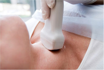
An ultrasound of the thyroid produces pictures of the thyroid gland and the adjacent structures in the neck. The thyroid gland is located in front of the neck just above the collar bones and is shaped like a butterfly, with one lobe on either side of the neck connected by a narrow band of tissue. It is one of nine endocrine glands located throughout the body that makes and sends hormones into the bloodstream.
The thyroid gland makes the thyroid hormone, which helps to regulate a variety of body functions including how fast the heart beats. It is very common for patchy areas or nodules to develop in the thyroid that may or may not be felt on the skin surface. About five to 10 percent of adults will have lumps in their thyroid that doctor can identify on an exam. These are called palpable nodules. Ultrasound is very sensitive and shows nodules that cannot be palpated. In some age groups, nodules are seen on ultrasound in as many as 70 percent of adults. The vast majority of these are benign regions of thyroid tissue that pose no health risk. The minority of these are true tumours of the thyroid and may require further diagnosis or treatment.
-
What are some common uses of the procedure?
An ultrasound of the thyroid is typically used:
• to determine if a lump in the neck is arising from the thyroid or an adjacent structure
• to analyze the appearance of thyroid nodules and determine if they are the more common benign nodule or if the nodule has features that require a biopsy
• to look for additional nodules in patients with one or more nodules felt on physical exam
• to see if a thyroid nodule has substantially grown over time
-
How should I prepare?
No special preparation is required. You may need to remove all clothing and jewellery in the area to be examined. You may be asked to wear a gown during the procedure.
-
How is the procedure performed?
For most ultrasound exams, you will be positioned lying face-up on an examination table that can be tilted or moved.
A pillow may be placed behind the shoulders to extend the area to be scanned for a thyroid ultrasound exam.
After you are positioned on the examination table, the sonographer will apply a warm water-based gel to the area of the body being studied. The gel will help the transducer make secure contact with the body and eliminate air pockets between the transducer and the skin that can block the sound waves from passing into your body. The transducer is placed on the body and moved back and forth over the area of interest until the desired images are captured.
There is usually no discomfort from pressure as the transducer is pressed against the area being examined. However, if scanning is performed over an area of tenderness, you may feel pressure or minor pain from the transducer.
Once the imaging is complete, the clear ultrasound gel will be wiped off your skin.
An ultrasound of the thyroid is usually completed within 15-20 minutes.
-
Who interprets the results and how do I get them?
A radiologist, a physician specifically trained to supervise and interpret radiology examinations, will analyze the images and send a signed report to your primary care physician, or to the physician who requested the exam, and he/she will share the results with you.
Follow-up examinations may be necessary in some cases, and your doctor will explain the exact reason why another exam is requested. Sometimes a follow-up exam is done because a suspicious or questionable finding needs clarification with additional views or a special imaging technique. A follow-up examination may also be necessary so that any change in a known abnormality can be monitored over time. Follow-up examinations are sometimes the best way to see if treatment is working or if an abnormality is stable over time.
-
What are the benefits vs. risks?
Benefits
• Most ultrasound scanning is non-invasive (no needles or injections).
• Occasionally, an ultrasound exam may be temporarily uncomfortable, but it is almost never painful.
• Ultrasound is widely available, easy-to-use and less expensive than other imaging methods.
• Ultrasound imaging is extremely safe and does not use any ionizing radiation.
• Ultrasound scanning gives a clear picture of soft tissues that do not show up well on x-ray images.
• Ultrasound provides real-time imaging, making it a good tool for guiding minimally invasive procedures such as needle biopsies and fluid aspiration.
Risks
For standard diagnostic ultrasound, there are no known harmful effects on humans.
-
What are the limitations of an Ultrasound of the Thyroid?
If one or more nodules are detected within the thyroid gland, the radiologist will examine the features of the nodules. Some features are strongly suggestive that a nodule is benign in nature, and some raise concern that the nodule may be a true tumour. In other cases, the radiologist cannot distinguish between benign and malignant lumps with complete certainty. A fine needle biopsy and review of tissue under a microscope may be recommended for further evaluation, but in some cases surveillance and a repeat sonogram after a few months looking for stability may suffice.
It is not possible to determine thyroid function—that is, whether the thyroid gland is underactive, overactive, or normal—with ultrasound. For that determination, your doctor may order a blood test or a radioactive iodine uptake test.
Transrectal Ultrasound
 A transrectal ultrasound is an ultrasound technique that is used to view a man’s prostate and surrounding tissues. The ultrasound transducer (probe) sends sound waves through the wall of the rectum into the prostate gland, which is located directly in front of the rectum. Transrectal ultrasound may also be called prostate sonogram or endorectal ultrasound.
-
Why transrectal ultrasound is done
• determine if the prostate is enlarged
• diagnose prostate cancer
• Transrectal ultrasound is done if the doctor suspects cancer because of an increased prostate-specific antigen (PSA) level, abnormalities felt during digital rectal examination (DRE) or certain symptoms are present, such as problems with urination.
• help guide the doctor when taking biopsy samples of the prostate
• help diagnose the cause of infertility in a man
• help guide the doctor during treatments for prostate cancer
• Transrectal ultrasound is used to guide the placement of implants during brachytherapy.
• It may help the doctor perform cryosurgery.
• Transrectal ultrasound may be used to help deliver high-intensity focused ultrasound (HIFU).
-
How transrectal ultrasound is done
A transrectal ultrasound is usually done as on outpatient procedure in a doctor’s office, clinic or hospital. The test usually takes 15–30 minutes.
-
How do I prepare?
Some special preparation is needed before a transrectal ultrasound:
• Avoid taking Aspirin and other medications that may thin the blood for 7–10 days before the test.
• An enema is used 1–4 hours before the procedure to help clean out the colon.
• The man may be told to urinate to empty his bladder just before the procedure.
-
How the test is done?
You will be asked to wear a gown during the procedure. Patient usually lies on the left side with the knees bent toward the chest. A protective cover and lubricant are put on the ultrasound transducer (probe), then it is placed into the rectum. You may feel pressure or have a sensation of fullness in the rectum when the transducer is in place.
The transducer directs high-frequency sound waves at the prostate. A computer analyzes the echoes created by the sound waves and displays an image of the prostate gland on a monitor. The image shows how big the prostate is and whether there are any abnormalities.
-
What the results mean
Abnormal findings may include:
• enlarged prostate (benign prostatic hyperplasia)
• inflamed or infected prostate (prostatitis)
• prostate cancer
What happens if a change or abnormality is found:
The doctor will decide whether further tests, procedures, follow-up care or additional treatment is needed.
Venous Ultrasound
 Ultrasound is safe and painless, and produces pictures of the inside of the body using sound waves.
Venous ultrasound provides pictures of the veins throughout the body. Usually the venous ultrasound is performed for lower or upper extremities.
A Doppler ultrasound study may be part of a venous ultrasound examination.
Doppler ultrasound is a special ultrasound technique that evaluates blood flow through a blood vessel, including the body’s major arteries and veins in the abdomen, arms, legs, neck and head (in infants and children).
-
What are some common uses of the procedure?
The most common reason for a venous ultrasound exam is to search for blood clots, especially in the veins of the leg. This condition is often referred to as deep vein thrombosis or DVT. These clots may break off and pass into the lungs, where they can cause a dangerous condition called pulmonary embolism. If the blood clot in the leg is found early enough, treatment can be started to prevent it from passing to the lung.
A venous ultrasound study is also performed to:
• determine the cause of long-standing leg swelling. In people with a common condition called “varicose veins”, the valves that keep blood flowing back to the heart in the right direction may be damaged, and venous ultrasound can help the radiologist decide how best to deal with this condition.
• aid in the placement of a needle or catheter into a vein. Sonography can help locate the exact site of the vein and avoid complications, such as bleeding or damage to a nearby nerve or artery.
• map out the veins in the leg or arm so that pieces of vein may be removed and used to bypass a narrowed or blocked blood vessel. An example is using pieces of vein from the leg to surgically bypass narrowed heart (coronary) arteries.
• examine a blood vessel graft used for dialysis if it is not working as expected; for example, the graft may be narrowed or blocked.
If a line is placed in a vein of the legs or arms, there is a much higher chance of developing a clot around it due to the smaller vessel size (especially in infants and young children). In some instances, a clot can suddenly form in the arm due to compression of the vein at the inlet of the chest or in the left leg due to compression of the vein on the left side by an artery in the abdomen.
Doppler ultrasound images can help the physician to see and evaluate:
• blockages to blood flow (such as clots).
• narrowing of vessels.
• tumours and congenital vascular malformations.
-
How should I prepare?
You should wear comfortable, loose-fitting clothing for your ultrasound exam. You may need to remove all clothing and jewelry in the area to be examined.
You may be asked to wear a gown during the procedure.
A period of fasting is necessary only if you are to have an examination of veins in your abdomen (Abdominal aorta scan). In this case, you will probably be asked not to ingest any food or fluids except water for six to eight hours ahead of time. Otherwise, there is no other special preparation for a venous ultrasound.
-
How is the procedure performed?
For most ultrasound exams, you will be positioned lying face-up on an examination table that can be tilted or moved.
A clear water-based gel is applied to the area of the body being studied to help the transducer make secure contact with the body and eliminate air pockets between the transducer and the skin that can block the sound waves from passing into your body. The sonographer (ultrasound technologist) or radiologist then places the transducer on the skin in various locations, sweeping over the area of interest or angling the sound beam from a different location to better see an area of concern.
Doppler sonography is performed using the same transducer.
When the examination is complete, you may be asked to dress and wait while the ultrasound images are reviewed.
Single extremity ultrasound examination is usually completed within 20 minutes. More complex exams (bilateral extremities) may take a longer period of time.
-
Who interprets the results and how do I get them?
A radiologist, a physician specifically trained to supervise and interpret radiology examinations, will analyze the images and send a signed report to your primary care physician, or to the physician or other healthcare provider who requested the exam, and he/she will share the results with you.
Follow-up examinations may be necessary, and your doctor will explain the exact reason why another exam is requested. Sometimes a follow-up exam is done because a suspicious or questionable finding needs clarification with additional views or a special imaging technique. A follow-up examination may also be necessary so that any change in a known abnormality can be monitored over time. Follow-up examinations are sometimes the best way to see if treatment is working or if an abnormality is stable over time.
-
What are the benefits vs. risks?
Benefits
• Most ultrasound scanning is non-invasive (no needles or injections).
• Occasionally, an ultrasound exam may be temporarily uncomfortable, but it is almost never painful.
• Ultrasound is widely available, easy-to-use and less expensive than other imaging methods.
• Ultrasound imaging is extremely safe and does not use any ionizing radiation.
• Ultrasound scanning gives a clear picture of soft tissues that do not show up well on x-ray images.
• Venous ultrasound helps to detect blood clots in the veins of the legs before they become dislodged and pass to the lungs. It can also show the movement of blood within blood vessels.
• Compared to venography, which requires injecting contrast material into a vein, venous ultrasound is accurate for detecting blood clots in the veins of the thigh down to the knee. In the calf, because the veins become very small, ultrasound is less accurate. However, potentially dangerous venous clots are typically lodged in the larger veins.
Risks
For standard diagnostic ultrasound, there are no known harmful effects on humans.